Introduction: Studies have shown that tantalum (Ta) has more excellent bioactivity and stability than the clinical implant materials widely used, such as titanium, cobalt chromium alloy, hydroxyapatite coating[1]. It has been revealed that the material surface local microenviroment constructed of macro, meso, and micro structure determined the cell behaviors[2]. In order to further improve the bioactivity of tantalum, tantalum surface treatment has attracted more and more interests[3]. In this work, three types of nanostructure (Ta2O5 nanotube, nanocrater, F-contained nanocoating) on tantalum have been constructed by anodization, and the influence of these three nanostructures on primary osteoblast behaviors has been investigated in hope of offering useful reference to the development of the surface modification of tantalum implants.
Methods: Anodization was performed in a two-electrode configuration using a DC power supply. Tantalum sheets (99.95% purity, 10mm×10mm) were used as anode materials, and a platinum foil served as the cathode electrode. The electrolyte consisted of hydrofluoric acid (HF), H2O and DMSO in concentrated sulfuric acid (H2SO4). Nanotube, F-contained nanocoating and nano dimpled tantalum were fabricated at different voltage from 20-50V. All the samples were washed with distilled water followed by ultrasonication, and then dried by air stream. The surface physicochemical properties of these three nanostructures have been characterized by SEM, EDS, XRD, OCA, AFM, etc. The third generation of osteoblast extracted from SD rat was cultured on these there nanostructures. And the osteoblast behaviors have been characterized by SEM, fluorescent staining, cck-8, etc.
Results and Discussion: Highly ordered Ta2O5 nanotube arrays with 20-30 nm diameter, F-contained nanocoating (O: 67 at%, F: 16 at%, Ta: 17 at%) distributed with about 10 nm diameter nanoholes, and nano dimpled tantalum with 100-120 nm diameter (O: 10 at%, Ta: 90 at%) were in-situ prepared on the surface of pure Ta by anodization (see Fig. 1A-C). The contact angle of Ta2O5 nanotube arrays, F-contained nanocoating, and nanocrater was about 88o, 38o, 86o, respectively, and the surface roughness (RMS) was 37 nm, 343 nm, 82 nm. After 24 h incubation, osteoblasts on Ta2O5 nanotube arrays, nano dimpled tantalum spread fully, similar to the shape of polygon, but osteoblasts on F-contained nanocoating had long and narrow morphology (in Fig. 1D-F). The cck-8 results (in Fig. 2) showed, Ta2O5 nanotube, F-contained nanocoating and nano dimpled tantalum enhanced the adhesion and proheliformation of primary osteoblasts. The significant difference existed between Ta2O5 nanotube arrays and nano dimpled tantalum. Although the primary osteoblasts preferred to adhere on the Ta2O5 nanotube arrays compared with nano dimpled arrays at 12 h, the OD value on nano dimpled arrays was significant higher than Ta2O5 nanotube arrays at 7 days.
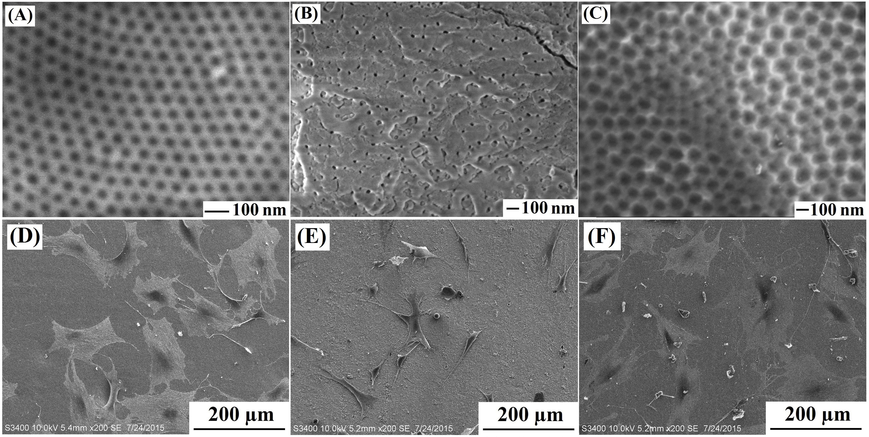
Fig. 1. SEM morphologies of nanostructure prepared by anodization: (a) Ta2O5 nanotube, (b) F-contained nanocoating, (c) nano dimpled tantalum; Cell SEM morphologies cultured on nanostructure showed above for 24 h: (D) Ta2O5 nanotube, (E) F-contained nanocoating, (F) nano dimpled tantalum.
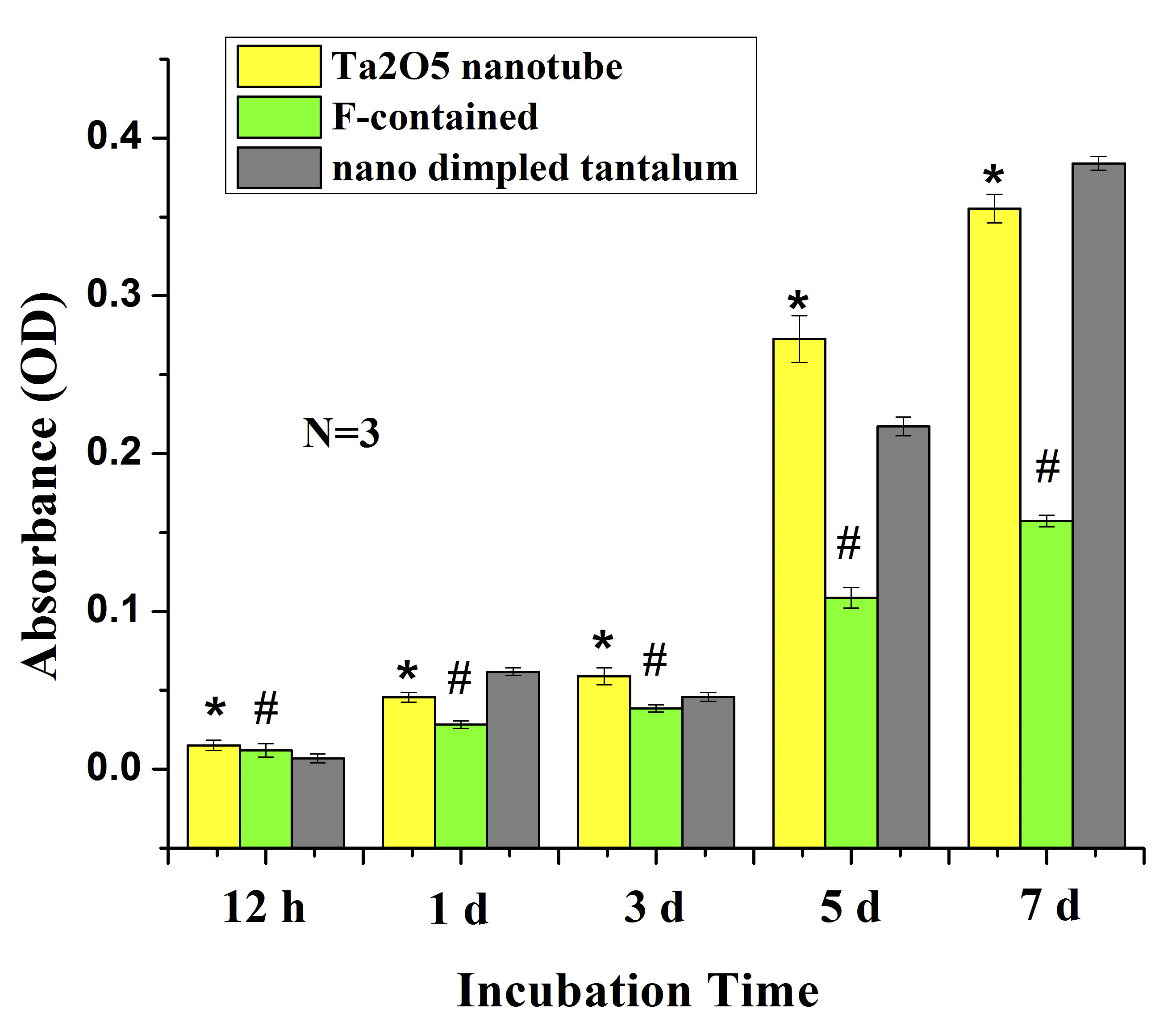
Fig. 2. Cell proliferation simulation according to cck-8 results. The bar graphs show mean±SD. * indicates a significant difference between Ta2O5 nanotube and F-contained nanocoating, nan dimpled tantalum (p<0.05), # shows a significant difference between F-contained nanocoating and nano dimpled tantalum.
Conclusions: In this work, Ta2O5 nanotube arrays, F-contained nanocoating, nano dimpled tantalum were fabricated by anodization on the surface of pure Ta. Their physicochemical properties and the influence on cell behaviors were investigated. Ta2O5 nanotube arrays and nano dimpled tantalum had higher contact angle and lower surface roughness than F-contained nanocoating. Although the primary osteoblasts preferred to adhere on the Ta2O5 nanotube arrays compared with nano dimpled arrays at 12 h, the nano dimpled arrays was more beneficial to osteoblasts proliferation than Ta2O5 nanotube arrays at 7 days. We believe our results will play useful reference role to the surface modification of tantalum implants.
National Natural Science Foundation of China (No. 51401126, No. 51271117, No. 51571142) and Shanghai Committee of Science and Technology, China (No. 14441901800)
References:
[1] M. Roy, V.K. Balla, S. Bose, A. Bandyopadhyay. Comparison of Tantalum and Hydroxyapatite Coatings on Titanium for Applications in Load Bearing Implants, Adv. Eng. Mater. 2010, 12: B637–B641.
[2] K. Anselme. Osteoblast adhesion on biomaterials, Biomaterials, 2000, 21: 667-681.
[3] Na Wang, Hongyi Li, Jinshu Wang, Su Chen, Yuanping Ma, Zhenting Zhang. Study on the anticorrosion, biocompatibility and osteoinductivity of tantalum decorated with tantalum oxide nanotube arrays films. ACS Applied Materials & Interfaces. 2012, 4: 4516-4523.