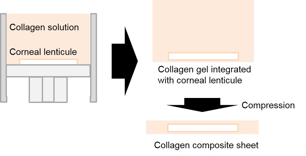
Introduction: The limbal epithelial stem cells (LESC), which is located in the basal epithelium of cornea, plays a crucial role of maintaining the integrity of the epithelium with their continuous population[1]. Thus, the failure of LESC can results in severe corneal disease accompanied by conjunctival epithelial ingrowth, neovascularization, inflammation and impaired vision. In this regard, collagen, which is main component of cornea, has been considered as a proper material to develop a LESC carrier to reconstruct the epithelium[2]. However, utilization of reconstituted collagen is restricted in in vitro structural remodeling due to insufficient mechanical property of collagen, and lenticule, decelluarized stromal tissue obtained from laser eye surgery, is also limited due to the poor cell attachment. In this study, collagen composite sheet with corneal lenticule and reconstituted collagen, which complementarily support deficiency in properties, was developed to enhance the mechanical property and cell attachment. Our approach of achieving the composite sheet is based on compression of reconstituted collagen gel containing the corneal lenticule[3]. Consequently, we can achieved the collagen composite sheet having good mechanical and cell attachment properties as the LESC carrier.
Materials and Methods: Type I collagen gel integrated with corneal lenticule was obtained by collagen neutralization process. The corneal lenticule was placed inside the customized mold filled with neutralized collagen solution followed by incubation under physiological condition. The composite gel was compressed to form a physically crosslinked composite sheet. The mechanical properties of the composite sheet was evaluated by spherical indentation test and in vitro enzymatic degradation tests. The limbal epithelial cell culture experiment was also performed to assess the cell attachment properties of the composite sheet.

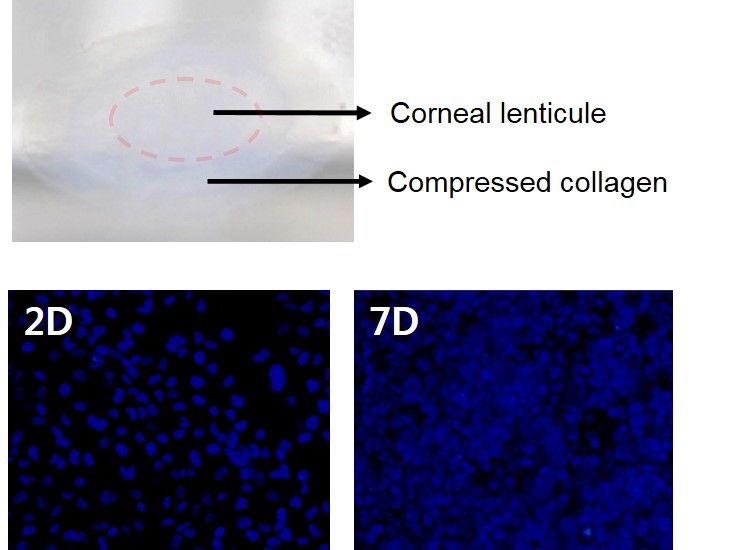
Results and Discussions: The corneal lenticule-reconstituted collagen composite sheet was successfully fabricated through gelation of type I collagen with the corneal lenticule and collagen compression process. The composite sheet shows more enhanced mechanical properties for both spherical indentation and degradation tests, which indicate that the composite sheet can be stably grafted onto the cornea and reliably support the cells during epithelium regeneration. The cell culture experiments results also showed that limbal epithelial cells can be stably attached and fully expanded on the sheet.

Conclusion: In this study, the collagen composite sheet was developed to overcome the limitations of conventional collagen based tissue equivalent for corneal epithelium regeneration. By integrating the corneal lenticule in the compressed collagen sheet, both mechanical and biological stabilities for delivering LESC can be obtained.
This work was supported by the National Research Foundation of Korea(NRF) grant funded by the Korea government(MSIP) (No. 2014R1A2A1A01006527); This work was supported by the Industrial Technology Innovation Program(No. 10048358) funded by the Ministry Of Trade, Industry & Energy(MI, Korea)
References:
[1] A. Shah, J. Brugnano, S. Sun, A. Vase, E. Orwin, “The Development of a Tissue-Engineered Cornea: Biomaterials and Culture Methods", Pediatr. Res., Vol. 63, 2008
[2] R. A. Brown, M. Wiseman, C. Chuo, U. Cheema, S. N. Nazhat, “Ultrarapid Engineering of Biomimetic Materials and Tissues: Fabrication of Nano- and Microstructures by Plastic Compression”, Adv. Funct. Mater., Vol. 15, 2005.
[3] Y. Oie, K. Nishida, “Regenerative Medicine for the Cornea”, Biomed. Res. Int., Vol. 2013, 2013.