Introduction: Peptide self-assembly has been widely explored for biomaterials applications, including cell delivery, drug delivery, and vaccine platforms. It has been previously demonstrated that epitope-bearing β-sheet fibrillizing peptides can elicit strong and specific antibody responses without supplemental immune adjuvants, making them an attractive platforms for vaccine development[1]. However, β-sheet fibrillizing peptides lack structural precision, and the kinetics of their assembly and disassembly are difficult to control. To address these shortcomings, we now report the design of a new self-assembly platform based on peptides that fold and assemble into α-helical nanofibers. This folding strategy allows for greater structural control and tunable rates of assembly and disassembly. We expect this control will be crucial in optimizing the materials’ trafficking and engagement of specific immune cells in vivo. The self-assembled peptide fiber was designed to incorporate both the universal CD4+ T cell epitope (PADRE, aKXVAAWTLKAa, where “X” is cyclohexylalanine, and “a” is D-alanine)[2] and a B-cell epitope (PEPvIII, LEEKKGNYVVTDH) targeting the epidermal growth factor receptor class III variant (EGFRvIII)[3], a tumor-specific receptor present in a significant proportion of glioblastomas and other human cancers. Both T cell and B cell epitopes were covalently linked to the helical peptide with C-terminal extensions. The antibody responses elicited by the epitope-bearing fiber were found by ELISA to be epitope-specific, dependent upon self-assembly, and comparable in titers to that raised by epitope peptide delivered in complete Freund’s adjuvant (CFA).
Materials and Methods: Peptides were synthesized as previously reported[1]. The morphology of the self-assembled fiber was studied using both circular dichroism (CD) and transmission electron microscopy (TEM). Immunizations were given in sterile PBS to female C57BL/6 mice subcutaneously. After 4 weeks, each mouse was boosted with one-half the primary dose. Mice in the CFA group were boosted with incomplete Freund’s adjuvant (IFA). Antibody titers were determined using ELISA.
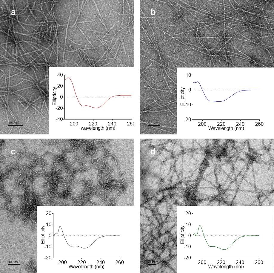
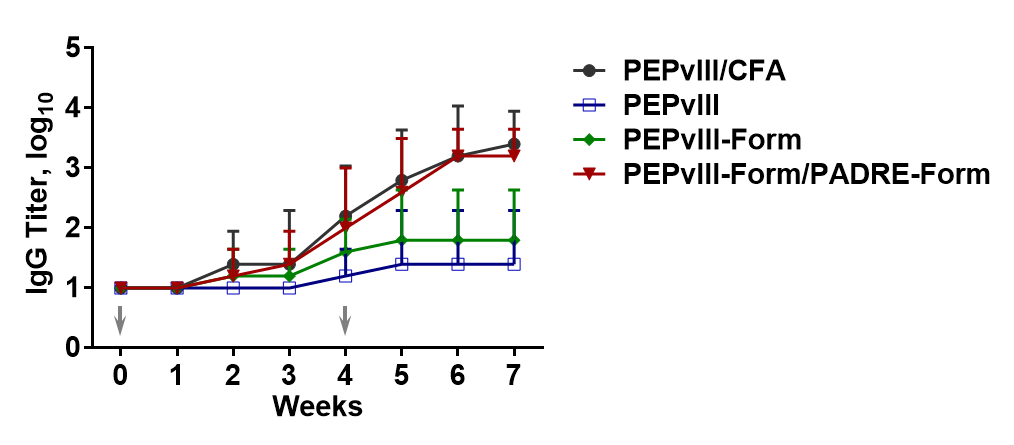
Results and Discussion: The Form-I peptide developed by Conticello and co-workers (QARILEADAEILRAYARILEAHAEILRAQ)[4] self-assembled into cylindrical α-helical fibers in our hands. With N-terminal extension of the epitope sequence, the modified Form peptide could still self-assemble into α-helical fibers, regardless of the hydrophobicity of the epitopes, according to TEM and CD. (Fig. 1) Although the morphology of PADRE-Form peptide appeared to be different (Fig. 1 c), the peptide fiber formed with PEPvIII-Form (Fig. 1 b) and mixing PEPvIII-Form and PADRE-Form (Fig. 1 d) both exhibited similar morphology to that of the Form peptide fiber (Fig. 1 a). The immunization study showed that PEPvIII peptide did not elicit strong antibody responses without being conjugated to the fibrillizing peptide. (Fig. 2, PEPvIII group) While PEPvIII- specific was antibodies were raised in mice immunized with only PEPvIII-Form peptide to about titer 2 after boost (Fig. 2, PEPvIII-Form group), the antibody titer was significantly higher in the mice immunized with PEPvIII-Form/PADRE-Form (Fig. 2. PEPvIII-Form/PADRE-Form Group), comparable to the peptide delivered with CFA (Fig. 2, PEPvIII/CFA group). These results indicated that the immunogenicity of the materials relied upon both the self-assembling fibrillar peptide and also T cell help.

Fig. 1. The TEM images and CD graphs of a) Form peptide fiber, b) PEPvIII-Form peptide fiber, c) PADRE-Form peptide fiber, d) PEPvIII-Form/PADRE-Form peptide fiber

Fig. 2. Anti-PEPvIII antibody responses observed in different mice groups. Half of primary immunization dose was given to each mice as boost injections
Conclusion: Epitope-bearing Form peptides self-assembled into α-helical fibers, with morphologies similar to the unmodified peptide. Specific antibody responses against an EGF receptor epitope relevant to the treatment of glioblastoma were raised without adjuvant. Additionally, the co-assembly of PADRE-Form augmented the response to a level similar to mice receiving a strong adjuvant, CFA. These results indicate that this platform may be useful in the development of self-adjuvanting vaccines.
NIH/NIAID 5R01AI118182
References:
[1] Rudra, J. S.; Tian, Y. F.; Jung, J. P.; Collier, J. H. Proc. Natl. Acad. Sci. U.S.A. 2010, 107, 622–7
[2] Pompano, R. R.; Chen, J.; Verbus, E. A.; Han, H.; Fridman, A.; McNeely, T.; Collier, J. H.; Chong, A. S. Adv Healthc Mater 2014, 3, 1898–908
[3] Choi, B.; Archer, G.; Mitchell, D.; Heimberger, A.; McLendon, R.; Bigner, D.; Sampson, J. Brain Pathology 2009, 19, 713-23
[4] Egelman, E. H.; Xu, C.; DiMaio, F.; Magnotti, E.; Modlin, C.; Yu, X.; Wright, E.; Baker, D.; Conticello, V. P. Structure 2015, 23, 280–9