Introduction: In this study the possibility to compare two different approaches based on inorganic and organic bioactive signals used to obtain biomimetic chitosan scaffolds is reported. Nowadays, current tissue engineering strategies are focused on the restoration of pathologically altered tissue architecture by transplantation of cells in combination with supportive scaffolds and biomolecules. Recentrly, considerable attention has been given to chitosan (CS)-based materials and their applications in the field of orthopaedic tissue engineering. Interesting characteristics that makes chitosan suitable for this purpose are a minimal foreign body reaction, an intrinsic antibacterial nature, and the ability to be molded in various geometries and forms such as porous structures, suitable for cell ingrowth and osteoconduction.
Experimental Methods: Chitosan scaffolds were prepared by using a foaming method based on physical foaming combined with microwave curing[1]. The biomimetic scaffolds were obtained by using two different approaches based on inorganic and organic compounds, respectively. The first one was based by treating the scaffolds with a supersaturated SBF solution (5SBF); the treatment is able to create an hydroxyapatite coating on the surface and into the porous wall in few days (7days). The second approach is based by using a bioactive peptide of Bone Morphogenetic Protein (BMP-2). The 67-87 residue of the “knuckle” epitope of hBMP-2 (NSVNSKIPKACCVPTELSAI) was synthesized by microwave-assisted Fmoc solid phase peptide synthesis (SPPS). The physicochemical, morphological and mechanical properties of materials, before and after treatments, have been evaluated. Furthermore, biological analysis by using hMSC cells to estimate the effect of the two different biomimetic approaches on cellular behaviour were performed.
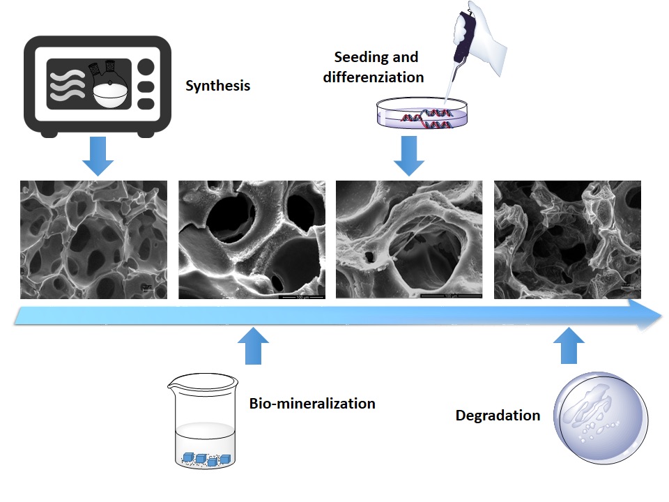
Results and Discussion: In this study, the combination of foaming method and biomimetic approaches has a great potential as process of production of chitosan based scaffold for bone tissue regeneration. The in vitro degradation tests showed a gradual dissolution of the foam over time, but maintaining their 3-D morphology and integrity even after 6 week of incubation. The surface morphology of the scaffolds after biomimetic treatment was observed using SEM-EDS analyses that demonstrated the presence apatite coating after few days of treatment. This data was confirmed by ATR-IR that also demonstrated the interactions between the polymeric matrix and peptide sequence. The kinetic release showed a sustained release in the time with a good effect on osteogenic differentiation of hMSC.The biological tests performed in basal medium without osteogenic factors demonstrated that the cell proliferation increases with time during the in vitro culture period suggesting that the materials are nontoxic, not affecting the cell proliferation and showed good biocompatibility. Moreover, for scaffold materials after inorganic and organic biomimetic approaches, an early osteogenic differentiation at 3 days was observed, demonstrating that a layer of apatite and/or BMP-2 peptide are bioactive solid signals for cells[2].
Conclusions: This study has demonstrated that the presence of bioactive signals on the scaffold surface allows to obtain an osteoinductive effect on hMSC, making chitosan a promising candidate scaffold material for bone tissue regeneration.
The authors acknowledge the Italian Ministry of Education, University and Research for providing partial financial support. The authors also thank Mrs. Cristina Del Barone for facilitating microscopy analysis.
References:
[1] C. Demitri, A. Giuri, M. G. Raucci, D. Giugliano, M. Madaghiele, A. Sannino, et al., "Preparation and characterization of cellulose-based foams via microwave curing," Interface Focus, vol. 4, February 6, 2014 2014.
[2] M. G. Raucci, M. A. Alvarez-Perez, C. Demitri, A. Sannino, and L. Ambrosio, "Proliferation and osteoblastic differentiation of hMSCs on cellulose-based hydrogels," J Appl Biomater Funct Mater, vol. 10, pp. 302-307, 2012.