Due to the increasing amount of implant requirements worldwide, there is a need to improve their sustainability and longevity to avoid potential replacements for implant recipients. An ideal implant is able to integrate with the natural bone tissue seamlessly without any complications. Modifications to the topography and chemistry of implant surfaces, such as titanium, can improve the osseointegration capabilities of the material[1]. An unconventional surface modification technique is the use of laser ablation to increase the roughness of the material to promote osseointegration.
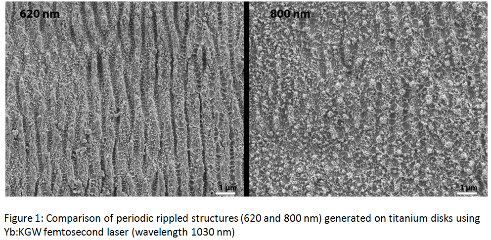
This study used a Yb:KGW femtosecond laser (Pharos from LightConversion), which when pulsed at 300 fs, was able to generate a ripple patterns on the surface of commercially pure grade 2 titanium disks (diameter of 15 mm) without the presence of cracks and craters typically found in laser modification techniques[2]. Specific ripple periodicities were generated with the laser (400, 620 and 800 nm) which correspond to, approximately, the length of 8, 12 and 16 units of collagen fibrils creating a semi-biomimetic surface. The laser ablation created a dual-topography on the surface, identified under SEM (Shown in Figure 1), consisting of the ripples along with resolidified molten droplets. Changes to the naturally occurring titanium oxide layer, which is known to promote osseointegration, were characterized using both auger electron spectroscopy and x-ray diffraction.
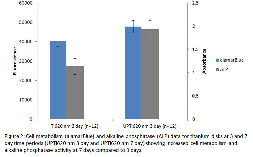
Saos-2, osteosarcoma, cells were seeded on to the surfaces of the titanium disks and allowed to grow for 3, 7 and 14 day time points to model mature human osteoblast behavior[3]. Cell metabolism and alkaline phosphatase activity were measured using alamarBlue and ALP assays respectively. Cell metabolism and ALP activity both showed statistically significant improvement on the laser ablated surfaces at early time compared to unmodified controls (Figure 2). The spread of the adhered cells was examined using fluorescent and scanning electron microscopy.
The results of this study show that the presence of this dual-topography was able to improve cell metabolism and ALP activity of osteoblast-like cells compared to controls. Additionally, the controlled ablations made with the Yb:KGW laser increased the roughness of the surface without any alterations to the chemical composition of titanium. Combined with the ease of manufacturing, these results promote the use of femtosecond laser-modified Ti as a biomaterial.
References:
[1] M. Geetha, a. K. Singh, R. Asokamani, and a. K. Gogia, “Ti based biomaterials, the ultimate choice for orthopaedic implants – A review,” Prog. Mater. Sci., vol. 54, no. 3, pp. 397–425, May 2009.
[2] A. Weck, T. H. R. Crawford, D. S. Wilkinson, H. K. Haugen, and J. S. Preston, “Laser drilling of high aspect ratio holes in copper with femtosecond, picosecond and nanosecond pulses,” Appl. Phys. A Mater. Sci. Process., vol. 90, no. 3, pp. 537–543, 2008.
[3] C. Pautke, M. Schieker, T. Tischer, A. Kolk, P. Neth, W. Mutschler, and S. Milz, “Characterization of Osteosarcoma Cell Lines MG-63 , Saos-2 and U-2 OS in Comparison to Human Osteoblasts,” Anticancer Res., vol. 24, no. 6, pp. 3743–3748, 2004.