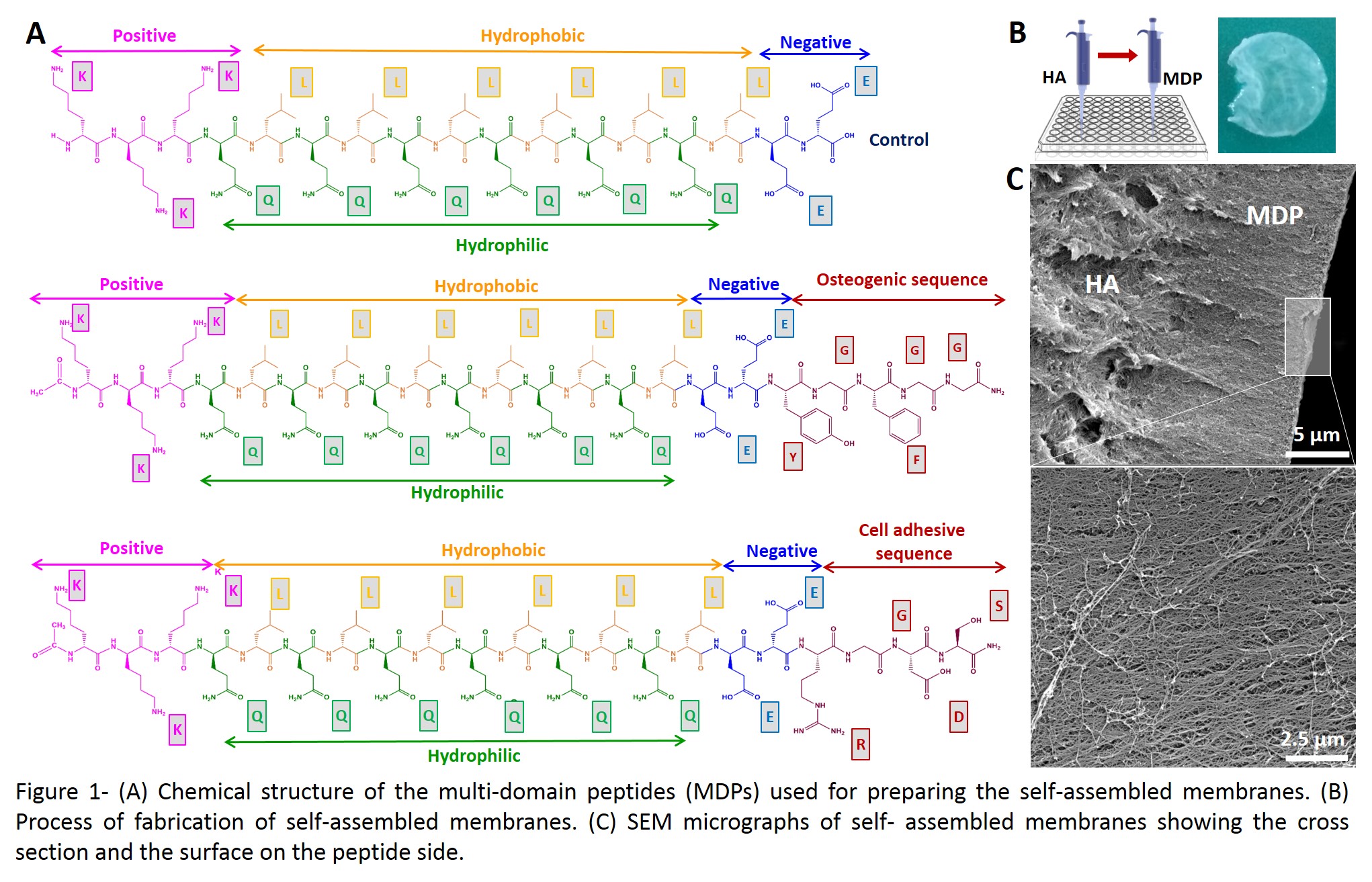
Introduction: Peptide self-assembly offers the possibility of fabricating highly organized biomaterials with the ability of recreating the nano-architecture and functionality of the extracellular matrix of tissues. The self-assembly of peptides with the negatively charged biopolymer hyaluronan (HA) has been explored to fabricate membranes with different biochemical and physical features[1]-[3]. Periosteum consists of microvascularized connective tissue covering the outer surface of cortical bone formed by two distinct layers: an outer fibrous layer that contains fibroblasts and Sharpey’s fibers; an inner layer called cambium, which contains multipotent mesenchymal stem cells and osteoprogenitor cells that contribute to normal bone growth, healing, and regeneration. To mimic the structure and function of periosteum, we designed new multi-domain peptides (MPDs) known to form filamentous nano-structures by self-assembly[4], to fabricate bilayer, periosteum-like, membranes by self-assembly with HA.
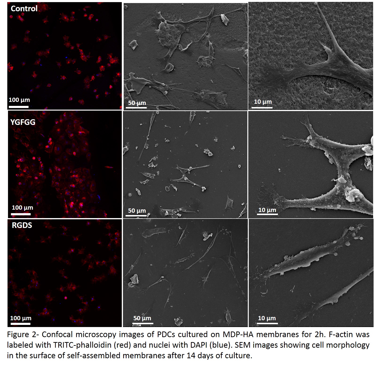
Materials and Methods: MPDs used in this study are composed of a positively charged block (to interact with HA), a β-sheet forming segment and bioactive domain aiming to promote cell adhesion (RGDS), mineralization (EE) and osteogenic differentiation (YGFGG) (Fig.1-A). The peptides were synthesised by solid phase and purified as described elsewhere[3]. Their secondary structure and charge were analysed by circular dichroism and zeta-potential, respectively. Membranes were obtained by interfacial self-assembly between aqueous solutions of HA (700 kDa, 2%) and the various MPDs (3%) (Fig.1-B). The microstructure of the membranes was analysed by scanning electron microscopy (SEM) and a preliminary cell adhesion assay was performed using periosteum-derived cells (PDCs) to assess their ability to support cell adhesion in the absence of serum. The cells were seeded on top of the membranes and cultured for 24 h. To examine the morphology of adherent cells, cells were first stained with TRITC-conjugated phalloidin and DAPI and then observed under a confocal microscope. For cell proliferation studies, PDCs were cultured with serum for 14 days. The samples were fixed and dehydrated for observation by SEM.
Results and Discussion: All designed peptides were successfully synthesised, maintaining the ability to form beta-sheet secondary structures. The HA-peptide membranes show a well-defined stratified organization with a thickness of ∼55 μm (Fig.1-C, cross-section) and a nanofibrillar structure. The presence of RGDS clearly enhances the adhesion of PDCs, when compared with control membranes (Fig.2). After 14 days of culture, a large number of well-spread cells with an elongated morphology are observed adhered to the membrane.
Conclusions: We anticipate that these highly organized biomimetic membranes can provide biochemical signals for stem cell growth and differentiation and be used to promote bone regeneration.
This work was supported by the Portuguese Foundation for Science and Technology under the scope of the project PTDC/CTM-BIO/0814/2012 and by the European Regional Development Fund (ERDF) through the Operational Competitiveness Programme “COMPETE” (FCOMP-01-0124-FEDER-028491).
References:
[1] D. S. Ferreira et al. “Hyaluronan and self-assembling peptides as building blocks to reconstruct the extracellular environment in skin tissue”, Biomaterials Science. vol. 1, 952-964, 2013.
[2] D. S. Ferreira et al. “Molecularly Engineered Self-Assembling Membranes for Cell-Mediated Degradation”, Advanced Healthcare Materials. vol. 4, 602–612, 2015.
[3] A. C. Mendes et al. “Co-Assembled and Microfabricated Bioactive Membranes”, Advanced Functional Materials. vol. 23, 430-438, 2013.
[4] H. Dong et al. “Self-Assembly of Multidomain Peptides: Balancing Molecular Frustration Controls Conformation and Nanostructure”, J. Am. Chem. Soc. vol. 129, 12468-12472, 2007.