Introduction: Spatiotemporally controlled delivery of biomolecules such as growth factors and antibiotics has been proposed as an alternative to systemic delivery[1]. Fibrous silk membranes have been used to obtain such controlled and sustained delivery of biomolecules[2]. However, release of drugs from silk is hard to control due to lack of interactions between most biomolecules and silk. Gelatin nanospheres (GNs), on the other hand, are suitable vehicles for the controlled delivery of a variety of biomolecules due to their charged nature[3],[4]. Therefore, we have focused on the synthesis of silk-based drug release system containing oppositely charged GNs to improve control over the delivery of multiple bioactive molecules. To this end, we developed a simple core-shell electrospinning technique to fabricate silk nanofibers containing homogeneously distributed and oppositely charged GNs.
Materials and Methods: Positively charged GNs (GANs, made of Gelatin Type A) and negatively charged GNs (GBNs, made of Gelatin Type B) were prepared by a two-step desolvation method[3] and were fluorescently labeled in PBS with DyLight™ 405 and 650 NHS esters, respectively. Regenerated silk fibroin was extracted from silk cocoons according to a previously developed protocol[5]. Nanofibrous membranes containing GANs and GBNs were prepared by core-shell electrospinning of silk/poly (ethylene oxide) (PEO) (3:1, wt/wt) suspensions containing GANs or GBNs at different feeding rates between core and shell suspensions, while nanofibers containing only GANs or GBNs were prepared by single nozzle electrospinning. The presence and distribution of GNs inside the nanofibers were characterized by confocal laser scanning microscopy. Moxifloxacin and vancomycin were added into GANs and GBNs suspensions, respectively, prior to preparation of drug-loaded nanofibrous membranes using electrospinning. The release kinetics of moxifloxacin and vancomycin from nanofibrous membranes were monitored by high performance liquid chromatography.
Results and Discussion:
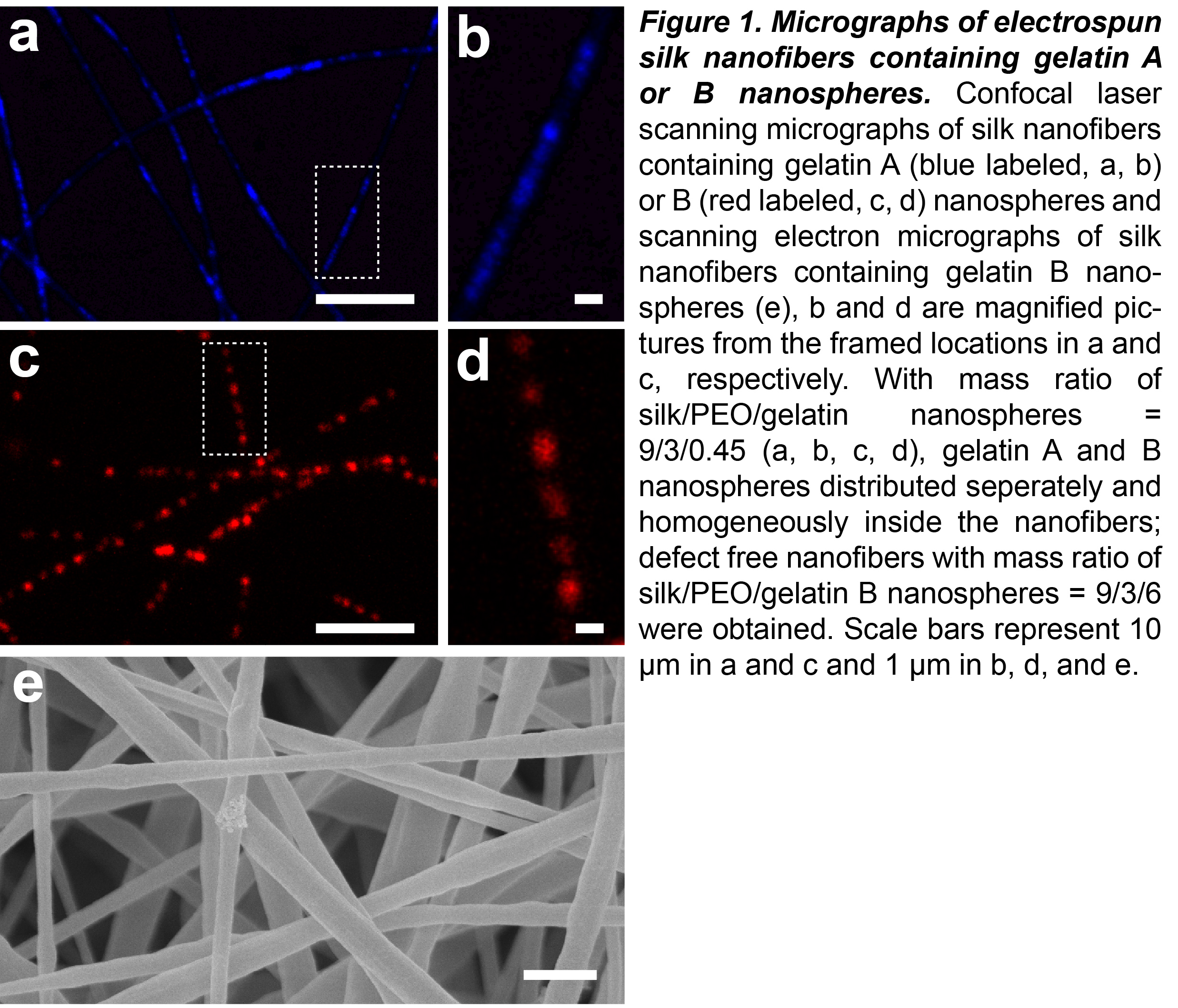
Suspensions containing approximately 33.3 wt% of GNs could be electrospun into nanofibers without any structural inconsistencies (Figure 1e). GANs or GBNs were homogeneously distributed inside the nanofibers (Figure 1a-d). Core-shell electrospun nanofibers (Figure 2f) exhibited more co-localization of GANs and GBNs than single nozzle electrospun nanofibers (Figure 2e), while single nozzle electrospinning obtained more dispersed single GANs or GBNs. Moreover, the ratio and distribution of GANs and GBNs inside the nanofibers could be fine tuned by adjusting the feeding rates of core and shell suspensions, which offers the opportunity for independent control over the delivery of multiple biomolecules.

Conclusion: Gelatin nanospheres were successfully incorporated into silk/PEO nanofibers to form fibrous “nano-in-nano” composites. The simple core-shell electrospinning technique can be utilized to fabricate nanofibers containing both GANs and GBNs, which enabled the fine tuning of the ratio and distribution of GANs and GBNs inside the nanofibers. The nanofibrous membranes can be used to delivery multiple biomolecules with adjustable release profile.
China Scholarrship Council (CSC), project number: 201206150058; NutsOhra Foundation, project number: 1303-024
References:
[1] Bongio M, van den Beucken JJJP, Leeuwenburgh SCG, Jansen JA. Development of bone substitute materials: from 'biocompatible' to 'instructive'. Journal of Materials Chemistry 2010;20:8747-59.
[2] Yucel T, Lovett ML, Kaplan DL. Silk-based biomaterials for sustained drug delivery. J Control Release 2014;190:381-97.
[3] Wang H, Zou Q, Boerman OC, Nijhuis AW, Jansen JA, Li Y, et al. Combined delivery of BMP-2 and bFGF from nanostructured colloidal gelatin gels and its effect on bone regeneration in vivo. J Control Release 2013;166:172-81.
[4] Song J, Odekerken JC, Lowik DW, Lopez-Perez PM, Welting TJ, Yang F, et al. Influence of the Molecular Weight and Charge of Antibiotics on Their Release Kinetics From Gelatin Nanospheres. Macromolecular bioscience 2015;15:901-11.
[5] Rockwood DN, Preda RC, Yucel T, Wang X, Lovett ML, Kaplan DL. Materials fabrication from Bombyx mori silk fibroin. Nature protocols 2011;6:1612-31.