Introduction: Microscopic particles with nanoparticles attached on the surface mimicking the surface morphology of viruses or raspberry have attracted much attention in recent years. However, the preparation methods are limited to electrostatic interaction or emulsion polymerization, most of which usually make use of silica or polystyrene particles as building materials[1]. The surface roughness is utilized to construct superhydrophobic surfaces, stabilize particles or enhance cellular delivery[2]. Yet, the influence of different roughnesses on cellular uptake has not been researched so far. Herein, a novel kind of porphyrin micro-nano particles is prepared through a newly-developed stimuli-responsive self-assembly method. Their roughness can be adjusted easily and are intended to be used in the research of effect of roughness on endocytosis.
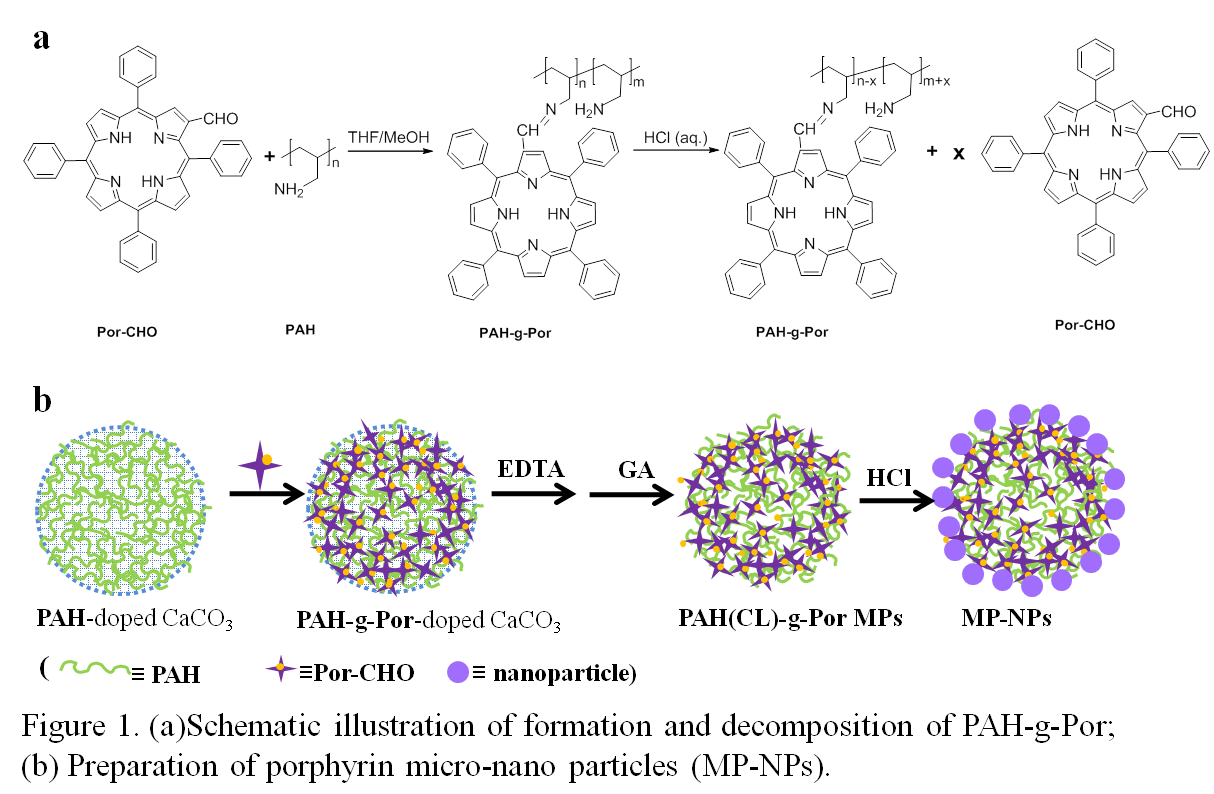
Materials and Methods: First, poly(allylamine hydrochloride) (PAH)-doped CaCO3 microparticles were reacted with 2-formyl-5,10,15,20-tetraphenylporphyrin (Por-CHO) by Schiff base formation. After template removal with EDTA and crosslinking with glutaraldehyde, the PAH-g-porphyrin microspheres (PAH-g-Por MPs) were obtained. Then the MPs were dispersed and incubated in pH 1 HCl to prepare the micro-nano particles (MP-NPs) (Figure 1). The roughness of MP-NPs was adjusted by the crosslinking degree of PAH-g-Por MPs and the incubation time in HCl.
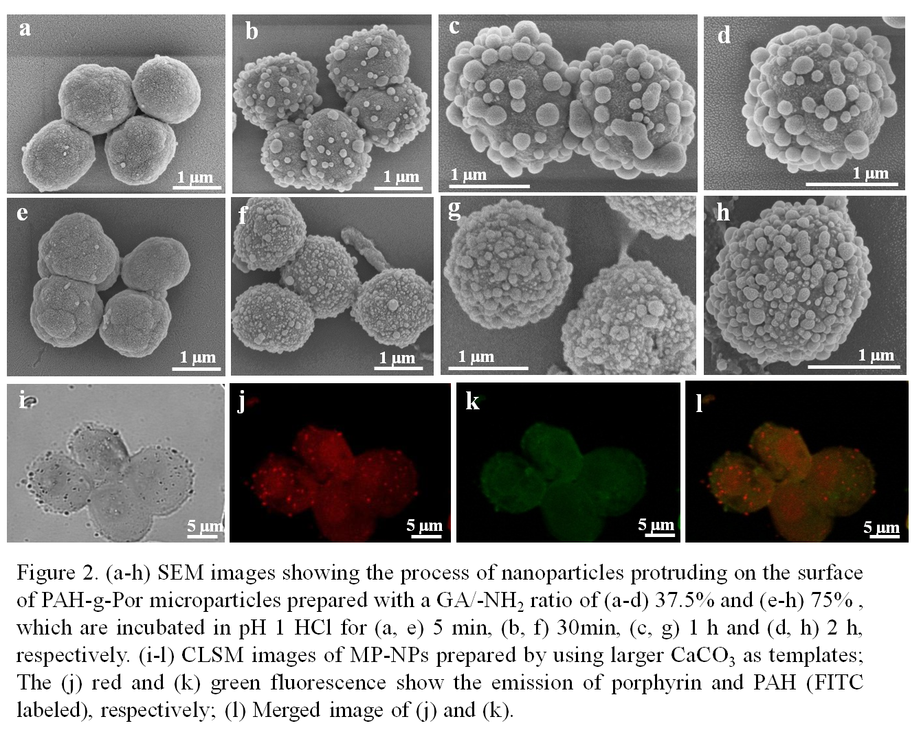
Results and Discussion: Spherical PAH-g-Por MPs with an average diameter of 1.5 μm were obtained, exhibiting a relatively smooth surface morphology regardless of different crosslinking degrees. After being incubated in HCl of pH 1, nanoparticles (NPs) were protruded on the surface of the MPs (Figure 2a-h). While the size of MPs had no obvious change, the size of NPs varied depending on the feeding ratio of GA. For the GA/-NH2 ratio (mol/mol) of 37.5%, the NPs became 185± 25 nm after 2 h (Figure 2d). However, for the 75%, the NPs were 105± 10 nm (Figure 2h). In the FITC-labeled MP-NPs, the NPs only showed the red fluorescence of Por but no green fluorescence of PAH-FITC, demonstrating their component was Por. Based on the previous work[3], the formation of MP-NPs should be attributed to the hydrolysis of partial Schiff base bonds triggered by low pH and the assembly of released Por-CHO. The original MPs prepared with a GA/-NH2 ratio of 75% were denser than those with 37.5%. As a result, the decomposition-assembly process was slower, resulting in smaller NPs on the surface.
Conclusion: A novel kind of porphyrin micro-nano particles (MP-NPs) mimicking the surface morphology of viruses is prepared with a pH-responsive self-assembly method. The size of the NPs can be adjusted by the crosslinking degree and the incubation time in HCl. The formation of MP-NPs results from the decomposition of Schiff base and self-assembly of Por. This method opens a new horizon for constructing virus-mimicking structures and the MP-NPs with different roughnesses are intended to be used for studying the influence of roughness on endoscytosis. Moreover, the NPs can be selectively modified to adjust the interaction between MP-NPs and cells by taking advantage of the aldehyde group of Por-CHO.
Financial supports by Ph. D. Programs Foundation of Ministry of Education of China (20110101130005), and the Natural Science Foundation of China (51120135001).
References:
[1] Agrawal, M.; Gupta, S.; Stamm, M. J. Mater. Chem. 2011, 21, 615–627
[2] Niu,Y. T.; Yu, M. H.; Yu, C. Z. et al. Adv. Mater. 2013, 25, 6233–6237
[3] Zhang, W. B.; Feng, Y. Q.; Gao, C. Y. et al. Langmuir 2015, 31, 4330−4340