In vivo evaluation of the role of carbonate ion in hydroxyapatite for bone remodeling
-
1
Institute of Biomaterials and Bioengineering, Tokyo Medical and Dental University, Materials Biofunction, Japan
-
2
Graduate school of Medical and Dental Sciences, Tokyo Medical and Dental University, Fixed Prosthodontics, Japan
-
3
Institute of Biomaterials and Bioengineering, Tokyo Medical and Dental University, Inorganic Biomaterials, Japan
Introduction: Recently artificial bone graft materials are widely available in dental and orthopedic surgery as an alternative to autogenous bone graft. Hydroxyapatite (HAp) is one of the most popular materials in medical application due to its superior biocompatibility and unlimited in supply. However, HAp is also reported to be no or only very sluggish biodegradation, which indicating that HAp prevents bone remodeling.
To control the degradation time, much attention has been paid to carbonated apatite (CA), which is nonstoichiometric HAp with carbonate ions substituting for phosphate ions. The carbonate substitutions in the apatite lattice are shown to increase the extent of solubility in weak acids, and bone remodeling.
Although biocompatibility of HAp and CA were assessed in vitro studies, due to the complex cell reactions around the artificial bone substitutes within an organism, animal experiments are widely used to evaluate the osteoconductivity or behavior of implanted materials. In this study, to evaluate the apatite porous block as bone substitutions, HAp and CA blocks were implanted into rabbit femurs and tibiae, and histologically analyzed.
Materials and Methods: HAp and CA powders were synthesized by a wet method. Porous apatite blocks were fabricated using these apatite powders. To control the porosity, the weights of paraffin beads were adjusted as 75% compared to apatite powders.
Japanese white male rabbits were used in this study (Approved by the animal experimental ethics committee of Tokyo Medical and Dental University (0110093A)). A bone defects were created approximately 3 mm in diameter in the medial epicondyle, and the inside facies medialis tibiae. At 4 and 12 weeks after implantation, the rabbit was euthanized and subjected to histological evaluation. The resin embedded samples were cut into 30 µm sections, and then stained using toluidine blue solution. Bone density, apatite density, and hard tissue density in the apatite block region were measured using micro-CT.
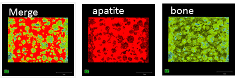
Results and Discussion: In this study, to analyze the bone formation and the behavior of apatite blocks, we separated the regions of bone tissue and porous apatite on the basis of a difference of radiolucency. The apatite densities analyzed by micro-CT were 28.4±2.4% (HAp), and 37.6±3.5% (CA), respectively. Although apatite densities of HAp were stable at 4 and 12 weeks after implantation, porous CA blocks showed bioresorbable properties.
Bone formation in the HAp blocks implanted into the cancellous bone was superior to that in the CA blocks. Although the bone density of HAp and CA blocks showed around 50% in the cortical regions after 4 weeks, due to the bioresorbable properties of CA blocks, bone formation in the CA blocks after 12 weeks increased and the defect was filled with bone tissues.
Conclusions: To substitute carbonate ion for phosphate ion increases the bioresorbable properties of porous apatite blocks and promotes bone remodeling.
Keywords:
Bone Regeneration,
in vivo,
Calcium phosphate,
Biodegradable material
Conference:
10th World Biomaterials Congress, Montréal, Canada, 17 May - 22 May, 2016.
Presentation Type:
Poster
Topic:
Biomaterials evaluation in animal models
Citation:
Nozaki
K,
Fujita
K,
Horiuchi
N,
Nakamura
M,
Miura
H,
Yamashita
K and
Nagai
A
(2016). In vivo evaluation of the role of carbonate ion in hydroxyapatite for bone remodeling.
Front. Bioeng. Biotechnol.
Conference Abstract:
10th World Biomaterials Congress.
doi: 10.3389/conf.FBIOE.2016.01.01129
Copyright:
The abstracts in this collection have not been subject to any Frontiers peer review or checks, and are not endorsed by Frontiers.
They are made available through the Frontiers publishing platform as a service to conference organizers and presenters.
The copyright in the individual abstracts is owned by the author of each abstract or his/her employer unless otherwise stated.
Each abstract, as well as the collection of abstracts, are published under a Creative Commons CC-BY 4.0 (attribution) licence (https://creativecommons.org/licenses/by/4.0/) and may thus be reproduced, translated, adapted and be the subject of derivative works provided the authors and Frontiers are attributed.
For Frontiers’ terms and conditions please see https://www.frontiersin.org/legal/terms-and-conditions.
Received:
27 Mar 2016;
Published Online:
30 Mar 2016.
*
Correspondence:
Dr. Kosuke Nozaki, Institute of Biomaterials and Bioengineering, Tokyo Medical and Dental University, Materials Biofunction, Tokyo, Japan, Email1
Dr. Kazuhisa Fujita, Institute of Biomaterials and Bioengineering, Tokyo Medical and Dental University, Materials Biofunction, Tokyo, Japan, Email2
Dr. Naohiro Horiuchi, Institute of Biomaterials and Bioengineering, Tokyo Medical and Dental University, Inorganic Biomaterials, Tokyo, Japan, Email3
Dr. Hiroyuki Miura, Graduate school of Medical and Dental Sciences, Tokyo Medical and Dental University, Fixed Prosthodontics, Tokyo, Japan, Email4
Dr. Kimihiro Yamashita, Institute of Biomaterials and Bioengineering, Tokyo Medical and Dental University, Inorganic Biomaterials, Tokyo, Japan, Email5
Dr. Akiko Nagai, Institute of Biomaterials and Bioengineering, Tokyo Medical and Dental University, Materials Biofunction, Tokyo, Japan, Email6