Introduction: Around 3.5 million new cancer cases and 1.7 million deaths per year in Europe make cancer a critical public health problem, regarding both early diagnosis and treatment. The design and development of novel nanosystems integrating advanced functionalized nano-sized core particles and active agents is an issue of intense research.
In spite of NPs around 100 nm in size have been widely studied to target tumors due to the passive targeting EPR effect, in the case of poorly permeable pancreatic adenocarcinomas, the use of small particles below 60 nm size improve the diffusion inside the tumor[1]. Single chain polymer NPs (SCPNs) have been studied in this work, due to their reduced size, and easily functionalization capability.
Materials and Methods: SCPNs based on polymethacrylic acid (PMAAc) (<50nm-sized) were prepared in mild conditions (r.t., water, absence of catalysts) starting from a copolymer of methacrylic acid and alkoxyamine derivatives. The intra-chain collapse was obtained by controlled addition of DTPA with aldehyde terminal groups as a cross linker.
Those particles were functionalized in one pot reaction with a radionucleide chelating agent and suitable synthetic somatostatin receptor targeting peptides by means of a covalent approach for targeted imaging. NODA was used as selective chelating agent for 67Ga to perform radio labelling for SPECT imaging.
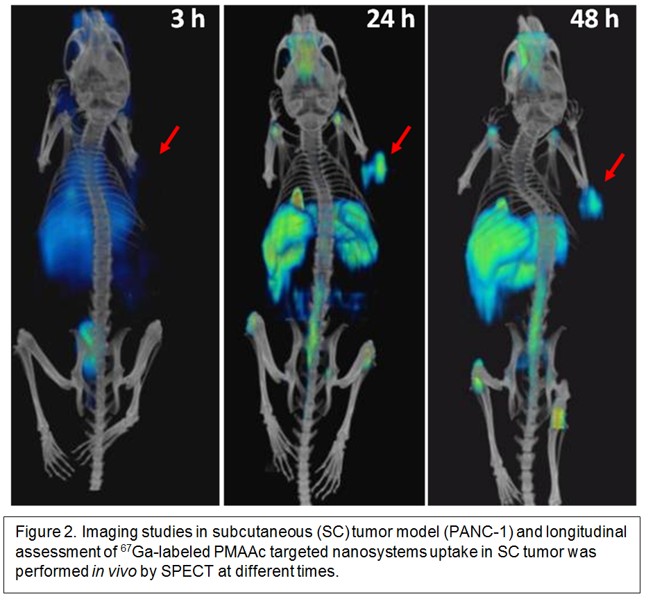
Cell viability was evaluated in vitro after 72 h of PMAAc-SCPNs incubation with six different cell lines by MTT assay. Acute toxicity screening after intravenous injection in male mice was performed by monitoring clinically, sacrifice after 24 h and hematology, clinical chemistry and histopathological examination. Imaging sessions of the pancreatic adenocarcinoma model in mice by SPECT/CT at 3, 24 and 48 h after injection were performed. Tumors were generated by subcutaneous injection of PANC-1 cells to male athymic-nude CD-1 Foxn1 nu/nu mice.
Results and Discussion: The collapse of the polymer chain for the formation of SCPNs was followed by DLS, 1H-NMR, GPC and TEM and yielded 16±5 nm (diameter) and a hydrodynamic diameter (Z-average) ~45nm (PDI 0.34) as determined by transmission electron microscopy (TEM) and dynamic light scattering (DLS).
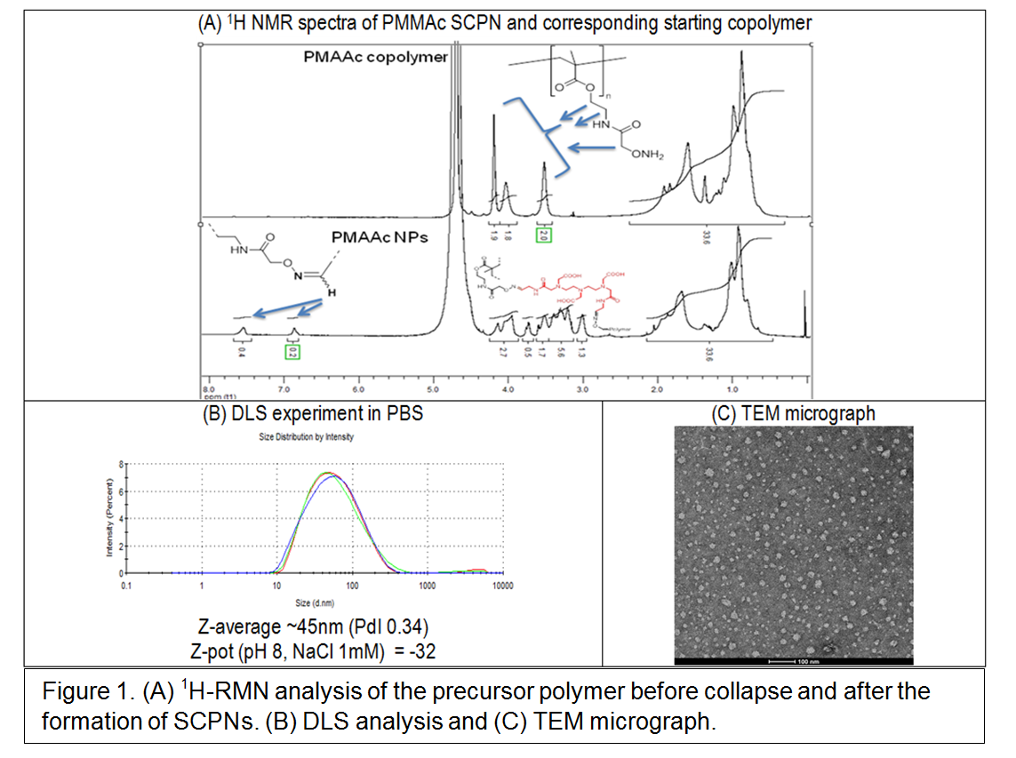
One pot amide coupling was carried out for the decoration of those particles with NODA and PTR-86 (selective targeting agent for somatostatin receptors). NODA amount was quantified by xylenol-orange test. 7% of fluorescein labeled PTR was anchored and quantified by fluorescence spectroscopy after purification by dialysis.
CD1 male mice were intravenously treated with a single i.v. dose of PMAAc SCPNs (12,5 and 100 mg/Kg) and sacrificed 24 h after treatment. Below 12.5 mg/kg the particles were safe. SPECT imaging showed significant signal increase tumor/muscle ratio when results at 24 and 48 hours were compared.
Conclusions: SCPNs showed interesting stability, biocompatibility and targeting properties once functionalized with PTR peptide. These SCPNs seem promising for further development as ongoing experiments are also demonstrating that they do not induce pro-metastatic side effects.
Save Me project has received funding from the European Union's 7FP for research, technological development and demonstration under grant agreement no. 263307.
References:
[1] S. Triantafyllos, J. Rakesh K., “Design considerations for nanotherapeutics in oncology”, Nanomedicine: Nanotechnology, Biology, and Medicine (2015), doi: 10.1016/j.nano.2015.07.015.