Introduction: In recent years, tissue regeneration by transplanting cardiomyocytes or various effector cells differentiated from induced pluripotent stem cells (iPS cells) or mesenchymal stem cells (MSC) are attracting great attentions. However, the fate of the transplanted stem cells including their distribution, migration, and even survival have not been clarified well. We have been focusing on the magnetic resonance imaging (MRI) for cell monitoring. Compared with optical tracking of the fluorescently labeled cells, MRI is remarkably advantageous in observation depth and resolution. In this study, novel water-soluble contrast agents were synthesized by introducing gadolinium (Gd) chelate (DOTA) to the terminal groups of branched poly(ethylene glycol) (PEG). Their longitudinal relaxation times (T1) and the contrast efficiency were evaluated, and the contrast agent with the best feature was subjected to the cell labeling and tracking.
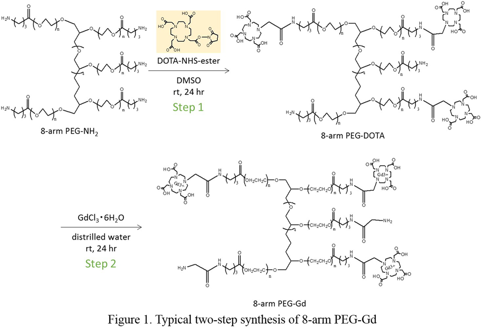
Experimental: Synthesis of PEG-Gd: PEG derivatives, 8-arm PEG-NH2 (Mn=15000), 4-arm PEG-NH2 (Mn=15000), dendron PEG-NH2 (Mn=40000), and linear PEG-NH2 (Mn=10000), were reacted with DOTA-NHS-ester in anhydrous dimethylsulfoxide (DMSO) at room temperature for one day, dialyzed with Spectra/Pore membrane (MWCO=3500) in distrilled water three times, and freeze-dried to obtain PEG-DOTA. PEG-DOTA was conjugated with GdCl3 to obtain PEG-Gd (Figure 1). The substitution ratio of DOTA and Gd conjugation were measured by proton nuclear magnetic resonance spectroscopy (1H-NMR) and inductively coupled plasma mass spectrometry (ICP-MS), respectively. T1 and MR images of each PEG-Gd aqueous solutions with different concentrations were measured by NMR and Magnetic resonance imaging (MRI, 1.5T).
iPS cells labeling by electroporation: The PEG-Gd was added to the iPS cell suspension in PBS, and electrical pulses were applied to the cell dispersions using a CUY-21 electroporator. The electrical pulses were optimized for efficient cell labeling. The cells were incubated for 10 min on ice bath and washed with PBS twice, then the labeling efficiency was measured by 1.5T MRI.
Results and Discussion: PEG-Gds having branched structure were found to show larger relaxivity than linear PEG-Gd and higher r1 than the commercial contrast agent irrespective of the Gd substitution ratio. According to the results of MRI, the luminance of each PEG-Gd is increased with the concentration increased. The 8-arm PEG-Gds having Gd conjugatoin of 46 % and 51% show the highest luminance suggesting the highest contrast efficiency. The in vitro cell labeling study also demonstrated that 8-arm PEG-Gd has the highest applicability.
Conclusion: Water-soluble PEG-Gd contrast agents with different structures were developed by two-step reaction. The relaxivity and luminance of the branched structure PEG-Gds were higher than linear PEG-Gd. The Gd introduction ration of 8-arm PEG-Gd did not affect the longitudinal relaxivity and luminance. The 8-arm PEG-Gd that showed much higher MR contrast than the commercially available contrast agent is the most efficient for cell tracking contrast agent.
JSPS KAKENHI Grant Number 15H06912