Introduction: Calcium-phosphate (CaP) nanoparticles decorated with silver (Ag) have shown potential to kill infectious bacteria in vitro.[1] [2]A coating that localizes the antibacterial CaP- Ag nanoparticles using osteoconductive chitosan may provide a means to inhibit bacterial biofilm formation and subsequent infectious complications for these implants. This work evaluated antimicrobial, cytocompatibility, and protein delivery properties of CaP- Ag in chitosan coatings.
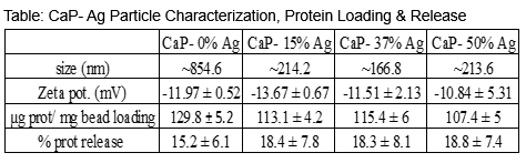
Materials and Methods: Porous CaP nanospheres with 0, 15, 37, or 50% Ag were fabricated using a hydrothermal synthesis of CaP followed by microwave heating of CaP-AgNO3 for Ag decoration on CaP. [1] Chitosan (82 DDA) coatings with 30wt% CaP-Ag particles were made via silane-solution casting on to cpTi and sterilized using ethylene oxide gas. Coated and uncoated cpTi coupons (n=3/group) were incubated with 3ml of bacteria (5X10⁶ colony forming units) under anaerobic conditions for common dental pathogens (P. gingivalis, ATC BAA-388; P. denticola, ATCC 35308) and aerobic conditions for orthopedic pathogens (P. aeruginosa: ATCC 15442; S. aureus, ATCC 25923, and S. epidermidis, ATCC 700576). Percent viability of bacteria was determined using MTT Bacterial Viability Assay (Roche Lab) using culture plastic controls. Coated and uncoated cpTi coupons were incubated with 2X10⁴ NIH 3T3 cells for 24 hrs and viability determined using CellTitre-Glo Assay (Promega). For protein release, 20mg of CaP-Ag particles (n=4/group) were incubated in 3mL of 1mg/mL α-chymotrypsin (MW = 25kDa, pI ~ 9.1), used as an analogue to BMP-2 (MW = 26kDa, pI ~8.2), for 24 hrs at 37C under gentle rocking. After 24hrs, supernatant was collected for determining protein loading by subtraction. Samples were incubated in 3mL PBS at 37C and eluates were collected with complete PBS every 2 days for 3 weeks. Protein concentration was determined using Quant-iT Protein Kit (Mol. Probe). Size and Zeta potential of particles was determined using Delsa™ Nano Submicron Particle Size and Zeta Potential Particle Analyzer (Beckmann Coulter).
Result:Uncoated cpTi, plain chitosan and chitosan coatings with CaP-0%Ag particles had minimal effect on viability of bacteria (Figure 1). There was a significant and dose-related decrease in viability of all bacteria with increasing Ag content on CaP nanoparticles with the CaP-50%Ag particles having the largest antibacterial properties (p<0.05). There were some differences in the sensitivity of bacteria to the coatings with S. epidermidis showing the highest sensitivity.
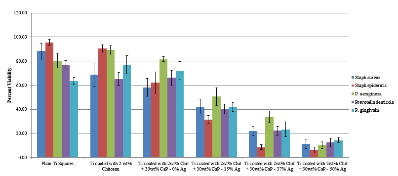
Figure1: Viability of bacteria on chitosan with CaP- Ag coatings
The total loading and percent release of protein was not different for the different CaP-Ag particles (Figure 2). This may be due to the similarly large negative Zeta potential of the particles (Table). Protein release pattern was also similar for the different CaP-Ag particles showing an initial burst release followed by a sustained release of approximately10 ng/µl from day 3 to 21.
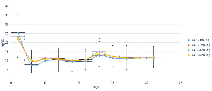
Figure2: Protein release from CaP- Ag particles in PBS
Discussion: There was no difference in the viability of the fibroblasts on the uncoated cpTi, plain chitosan and chitosan coatings with CaP-0%Ag particles. However, cell viability was reduced 100-fold on all chitosan coatings incorporating CaP-Ag particles regardless of the %Ag in the particles (data not shown). Part of reduced viability of the fibroblasts may be due to early release of Ag from particles due to the acetic acid used in the coating process. Previous studies showed that CaP-Ag particles alone had little effect on cell viability.[1]
Conclusion: Increasing concentration of Ag in CaP particles in chitosan coatings exhibited a dose dependent antibacterial effect on 5 implant pathogens in vitro. However, low cell viability to CaP-Ag particles in the coatings needs to be addressed. The CaP-Ag particles exhibited high protein loading and sustained protein release profile that may be advantageous for the local delivery of growth factors or other agents that require low and extended release profiles.
FedEx Institute of Technology, Memphis USA
References:
[1] Martin et al., SFB Mtng, Abst#767, April 2015
[2] Jennings, J.A., et al., Bacterial inhibition by chitosan coatings loaded with silver-decorated calcium phosphate microspheres. Thin Solid Films, 2015.