Introduction: Injectable hydrogels represent an useful strategy for drug delivery and tissue engineering as they can be easily placed into the defect site with minimum invasiveness[1]. Moreover, the introduction of nanoparticles in the polymer network can be a strategy to improve their general properties and influence their ability to act as carrier of biological factors[2]. Here we present the synthesis and characterization of a nanocomposite injectable hydrogel made of biocompatible polymers such as gelatin and chitosan in combination with nanodiamonds (ND)[3] as a carrier of the vascular endothelial growth factor (VEGF165) to promote angiogenesis in vitro.
Materials and Methods: Injectable hydrogels were prepared by combining chitosan (1.5% w/v) with beta-glycerophosphate (2% w/v) and gelatin (2% w/v) using genipin (0.02% w/v) as crosslinker. The ND suspension in water (0.05% w/v) with a dimension distribution of 20-60 nm was sonicated for 10 minutes prior mixing with the polymeric solution. Time sweep at 37°C along with frequency sweep studies in the range of 0.01 up to 10 Hz were carried out to investigate respectively the time of gelation and the influence of ND on the final strength. Young's elastic modulus was calculated from the slope of the curve stress strain in the range of 5-10% of strain. VEGF165 was complexed with ND suspension using a ratio ND/VEGF165 500:1 and the amount of VEGF165 loaded in each gel was 10 ng. Human umbilical vein endothelial cells (HUVECs) were seeded on the gels with a cell density of 50,000 cells and cultured in media without VEGF165 and bFGF. MTT assay to test ND cytotoxicity and actin and DAPI staining were assessed in vitro.
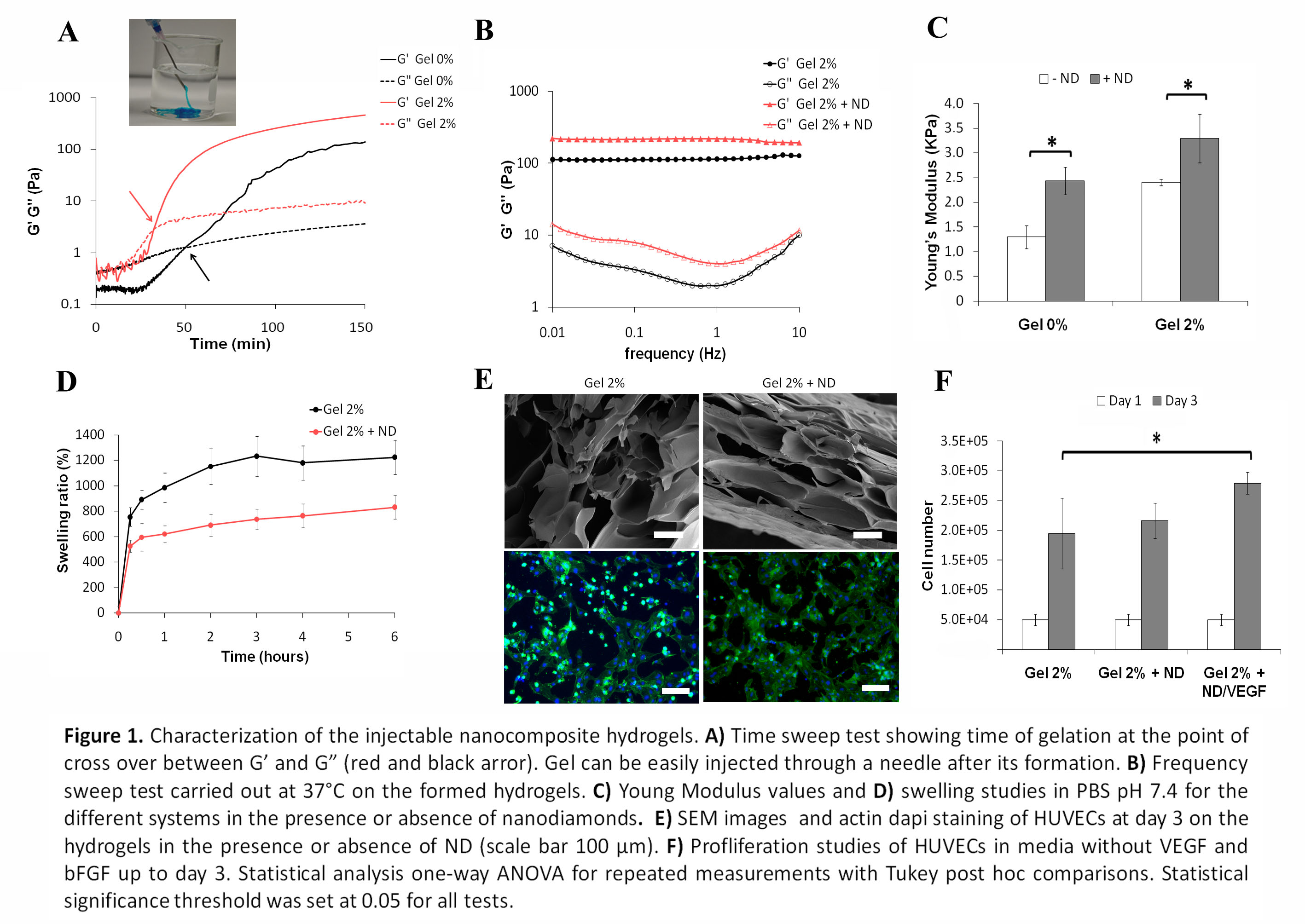
Results and Discussion: Genipin was kept constant in all formulations while the concentration of gelatin was varied up to 2% w/v (Gel 2%) and was found to reduce significantly the time of gelation (Fig 1A). Regarding the influence of ND on the gel mechanical properties, an increase in the value of G’ was detected in all formulations (Fig 1B) indicating a possible interaction of the ND with the polymeric network. The same trend was observed studying the Young’s modulus values obtained by compressing the hydrogels up to the point of breakage (Fig 1C). ND were able also to influence the swelling degree, reducing the amount of water uptaken by the hydrogels (Fig 1D) which can indirectly influence the release of loaded growth factors although no difference in porosity was observed among the different groups. Biocompatibility of the hydrogels were tested using HUVECs which were able to spread all over the surface at day 3 (Fig 1E) and a higher proliferation was observed when VEGF was loaded in the hydrogel (Fig 1F).
Conclusions: The presence of ND positively influenced the mechanical properties of the biocompatible injectable hydrogels. Morever ND were not cytotoxic and a higher proliferation of HUVECs was obtained when VEGF165 was loaded inside the gel in the presence of ND. Overall these results suggest the possible use of this injectable hydrogel to promote angiogenesis in vitro.
Arghya Paul likes to acknowledge the Institutional Development Award (IDeA) from the National Institute of General Medical Sciences, National Institutes of Health (NIH), under Award Number P20GM103638.
References:
[1] Bae KH, Wang L-S, Kurisawa M. Injectable biodegradable hydrogels: progress and challenges. Journal of Materials Chemistry B 2013;1:5371-88.
[2] Gaharwar AK, Peppas NA, Khademhosseini A. Nanocomposite hydrogels for biomedical applications. Biotechnology and bioengineering 2014;111:441-53.
[3] Mochalin VN, Shenderova O, Ho D, Gogotsi Y. The properties and applications of nanodiamonds. Nat Nano 2012;7:11-23.