Hydroxyapatite or calcite: How does physiological proteins influence the type, mechanism and kinetics of their formation on bioactive glasses?
-
1
The University of Manchester, School of Manchester, United Kingdom
-
2
Diamond Light Source, United Kingdom
-
3
University of Mons, Centre de Recherche en Modélisation Moléculaire, Belgium
-
4
STFC Rutherford Appleton Laboratory, Lasers for Science Facility, United Kingdom
-
5
University of Aston, School of Engineering & Applied Science, United Kingdom
-
6
Imperial College London, Department of Materials, United Kingdom
Introduction: Critical size bone defects, defined as those that do not heal completely through bone self-healing process, are becoming an increasing concern due to the prevalence of diseases as bone cancer, osteoporosis or trauma. One of the strategies to overcome these issues is to introduce temporarily a scaffold that mimics the extracellular matrix (ECM) and supports the natural bone self-healing process. To this effect, an adequate alternative is the use of biocompatible, bioactive and bioresorbable artificial materials as highly porous three dimensions (3-D) scaffolds[1]. Bioactive glasses are an attractive proposition due to their capability to produce a strong bond with bone tissue thanks to the formation of a hydroxyl carbonate apatite (HCA) layer under physiological conditions. The aims of this study are to investigate the influence of different physiological media on the type of surface reactions, their mechanism and kinetics of deposition of the mineral phase on the bioactive glass surface and how physiological proteins interact with these.
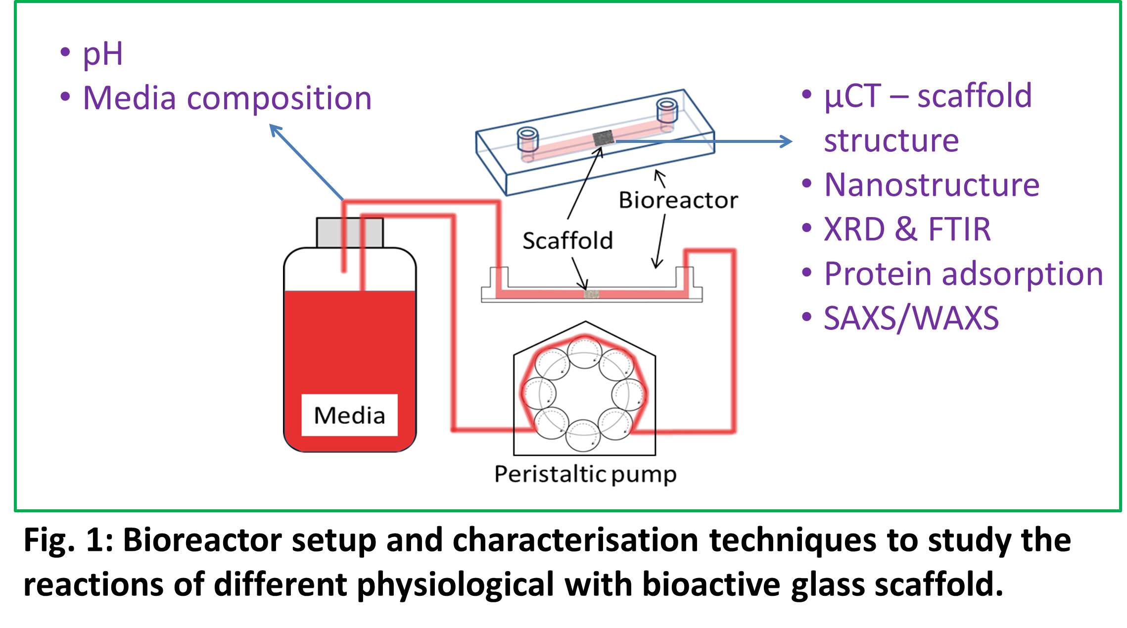
Material and Methods: Bioactive glass scaffolds were fabricated following the methods described in the literature[2],[3]. Scaffolds structure, chemistry and comspotion were characterised with several techniiques including synchrotron microtomography and simulataneous SAXS/WAXS. Surface reactions were performed using a flowcell as shown in Fig.1. Following reactions the scaffold and dissolution media were analysed using techqniues listed in Fig.1.
Results and Discussion: To our surprise we found that under flow conditions calcium carbonate precipitation dominated. It was oberved that within 20 minutes of flow peaks corresponding to calcite were visible. The presence of protein induced the calcium carbonate to grow in a particular orientation evidenced by a single sharp peak while the other peak intensities are almost null.
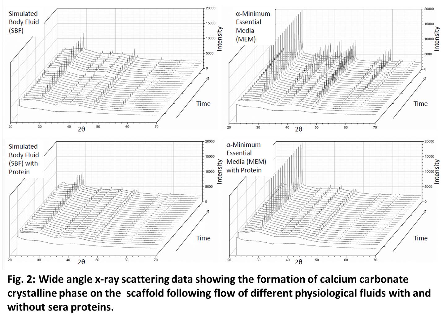
Figure 3a-c shows 3D and 2D images of the scaffold following the reaction in cell culture media. Almost all the surface were completely coated with a new phase, that was identified to be a polymorph of calcium carbonate. Figure 3 bottom shows competitive adsorption of albumin (green) and fibrinogen (red). It is clear from the images that the scaffold had a prefrential adsorption following preconditining in simulated body fluid and cell culture media.
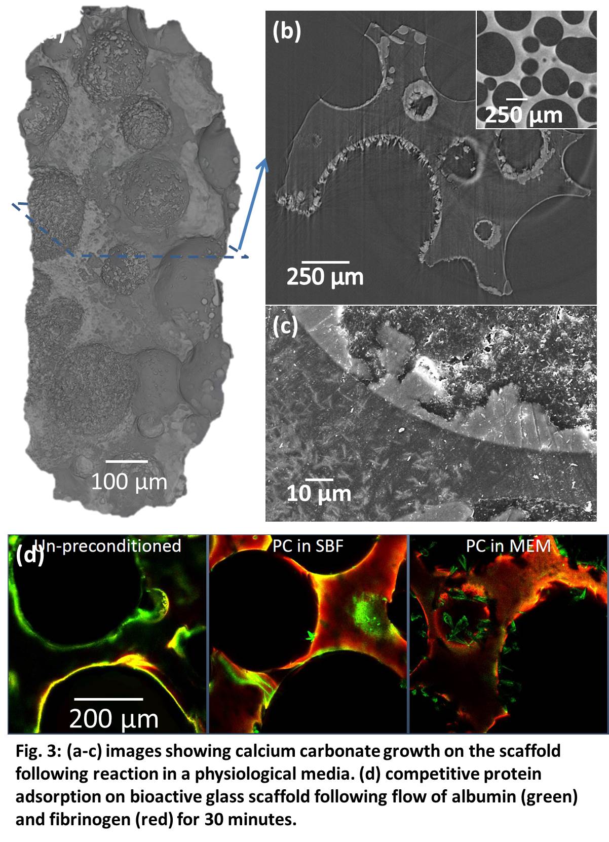
The results indicate that a calcium carbonate coating on the bioactive glass surface which has elicited a prefrential protein adsorption may be beneficial to bone growth. However, further in vivo experiments are required to confirm this.
Conclusions: Sol-gel derived bioactive glass under flow conditions formed a calcium carbonate phase on its surface. The calcium carbonate phase promoted the prefrential adsorption of albumin on to it while fibrinogen was found to adsorb on the scaffold surface. This suggests that calcium carbnate may have a role to play in the in vivo bone formatino on sol-gel derived bioactive glasses. However, in vivo tests are required to give a definite answer.
EPSRC (EP/I020861/1, and EP/I02249X/1, EP/M023877/1); Research Complex at Harwell; Diamond Light Source (SM11832-1); STFC, Central Laser Facility (App15130037)
References:
[1] J R Jones, Acta Biomater, 8, 4457-4486 (2013).
[2] P Sepulveda et al, J Biomed Mater Res, 59, 340-348 (2002)
[3] J R Jones et al, Biomaterials, 27, 964-974 (2006)
[4] FitzGerald et al, J Biomed Mater Res Part A. 91A(1), 76 (2009)
[5] S Midha et al, Acta Biomater, 9, 9169-9182 (2013)
Keywords:
protein,
Bioactivity,
Tissue Regeneration,
3D scaffold
Conference:
10th World Biomaterials Congress, Montréal, Canada, 17 May - 22 May, 2016.
Presentation Type:
New Frontier Oral
Topic:
Interfacial phenomena
Citation:
Poologasundarampillai
G,
Boix Alberich
M,
Clarke
D,
Smith
A,
Martin
R,
Lee
PD and
Jones
JR
(2016). Hydroxyapatite or calcite: How does physiological proteins influence the type, mechanism and kinetics of their formation on bioactive glasses?.
Front. Bioeng. Biotechnol.
Conference Abstract:
10th World Biomaterials Congress.
doi: 10.3389/conf.FBIOE.2016.01.02877
Copyright:
The abstracts in this collection have not been subject to any Frontiers peer review or checks, and are not endorsed by Frontiers.
They are made available through the Frontiers publishing platform as a service to conference organizers and presenters.
The copyright in the individual abstracts is owned by the author of each abstract or his/her employer unless otherwise stated.
Each abstract, as well as the collection of abstracts, are published under a Creative Commons CC-BY 4.0 (attribution) licence (https://creativecommons.org/licenses/by/4.0/) and may thus be reproduced, translated, adapted and be the subject of derivative works provided the authors and Frontiers are attributed.
For Frontiers’ terms and conditions please see https://www.frontiersin.org/legal/terms-and-conditions.
Received:
27 Mar 2016;
Published Online:
30 Mar 2016.