Introduction: In recent years, many of clinical reports have claimed that a chronic inflammatory process mediated by microglial cells is the fundamental process contributing to the death of dopamine (DA)-producing neurons owing to over generation of various cytotoxic mediators[1]. Also known as one of the most active polyphenol agents in green tea with antioxidant and anti-inflammatory effect, epigallocatechin-3-galate (EGCG) is potently able to suppress release of NO and TNF-α from LPS-induced microglial activation through the down-regulation of iNOS and TNF-α gene expression[2],[4]. Furthermore, the 67 kDa laminin receptor (67LR) also can interact with EGCG on surface of microglial cells as reported[3]. However, EGCG also has litmitations owning to its water soluble property and low bioavailability. Hence, the aim of this study is to design and develop a drug delivery system to overcome EGCG’s challenges. Interestingly, drug delivery systems based on polymeric carriers with potential advantages to deliver hydrophilic drugs are believed to easily overcome this challenges. In this stidy, poly(lactic-co-glycolic acid) (PLGA), a FDA-approved polymer, was fabricated as a microparticle system by double emulsion-solvent evaporation technique to sustain release of EGCG in the brain with excellent biocompatibility[5].
Materials and Methods: Epigallocatechin-3-galate (EGCG) was obtained from green tea by refluxing method. The property of radical scavenge by using 2,2-diphenyl-1-picrylhydrazyl (DPPH), cell viability and inhibitory effect of EGCG wrere carried out. EGCG loaded PLGA microparticles (EGCG-PLGA) were fabricated by double emulsion solvent evaporation technique, using H2O and poly(vinyl alcohol) (PVA) as hydrophilic phase and DCM as hydrophobic phase. Loading efficiency of EGCG, FTIR, SEM and upright micoroscopy were carried out. The release profile was carried out in PBS (pH 7.4) at 37 oC. Murine microglia cell lines (BV-2) were cultured in DMEM containing 10% FBS, 1% penicillin at 37 oC, 5% CO2 . The cell viability was determined by 3-[4,5-dimethylthiazol-2-yl]-2,5-diphenyl tetrazolium bromide (MTT) assay. Griess reagent was used to determine nitric oxide (NO).
Results and Discussion:
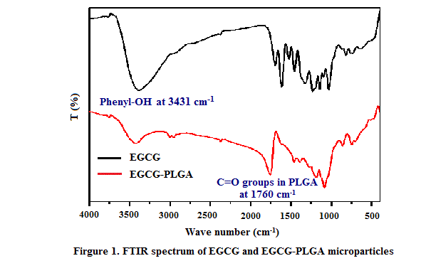
As can be observed in Figure 1, the spectrum of pure EGCG showed a sharp peak 3431 cm-1 indicating to phenyl-OH stretching. However, the intensity of this peak remarkably reduced after coated by PLGA microspheres in the spectra of EGCG-PLGA, which may be due to the influence of the large amount of PLGA coating. Besides, another sharp peak appeared at 1760 cm-1 considering that PLGA successful coated on EGCG surface.
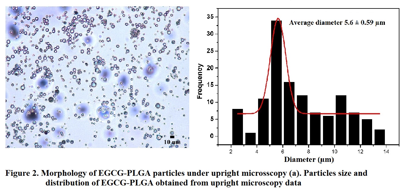
The result revealed that the morphology of EGCG-PLGA was spherical in shape, demonstrating the successful formation of spherical particles Figure 2(a). Particle size and distribution of EGCG-PLGA particles were determined in Figure 2(b). The data already showed the average particle size of EGCG-PLGA was 5.6 ± 0.59 µm, confirming the applicable ability of the material as a microparticle in drug delivery.
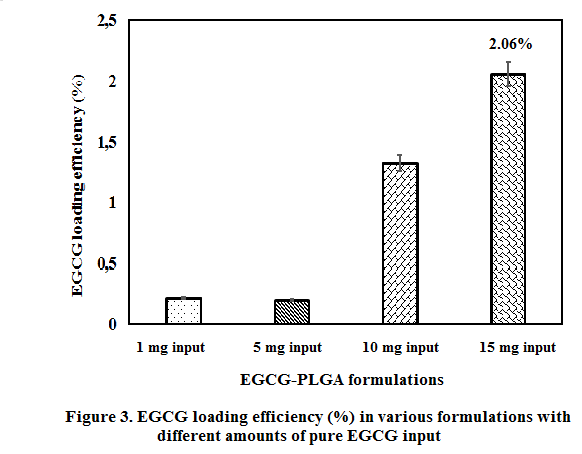
As described in Firgure 3, when increasing the EGCG loading amount, the EGCG encapsulation efficiency was raised and reached to 2.06%, aproximately 20.6 µg/mL EGCG per 1 mg/mL of material. This result revealed that EGCG-PLGA can be used for BV-2 microglial cells due to their low concentration that could not be toxic to cells. The results described that 1000 μg/mL of EGCG reached 90% scavenging activity of DPPH in 3 minutes, while 62.5 μg/mL of EGCG was unable to reach that efficacy even incubation time was extended. When LPS was administered, 50 μM of EGCG showed inhibitory effect on the activation of microgilas cells.
Conclusion: The results proved the formation and characterizarion of EGCG-PLGA microparticles, expressing a potential application of drug delivery system of this material to combat the inflammation reaction in microglial cells.
Ministry of Science and Technology, Taiwan (Grant No. 102-2632-M-033-001-MY3)
References:
[1] Qian, L. and P. Flood, Microglial cells and Parkinson’s disease. Immunologic Research, 2008. 41(3): p. 155-164.
[2] Li, R., et al., (−)-Epigallocatechin gallate inhibits lipopolysaccharide-induced microglial activation and protects against inflammation-mediated dopaminergic neuronal injury. Journal of Neuroscience Research, 2004. 78(5): p. 723-731.
[3] Fujimura, Y., et al., Green Tea Polyphenol EGCG Sensing Motif on the 67-kDa Laminin Receptor. Plos One, 2012. 7(5).
[4] Choi, D.K., S. Koppula, and K. Suk, Inhibitors of Microglial Neurotoxicity: Focus on Natural Products. Molecules, 2011. 16(2): p. 1021-1043.
[5] Danhier, F., et al., PLGA-based nanoparticles: An overview of biomedical applications. Journal of Controlled Release, 2012. 161(2): p. 505-522.