Collagen density gradient on poly(epsilon-caprolactone) surface for the design of 3D printed osteochondral
-
1
National Research Council of Italy, Institute of Polymers, Composites and Biomaterials, Italy
-
2
AO Research Institute Davos, Switzerland
-
3
National Research Council of Italy, Chemical Science & Materials Technology Department, Italy
Introduction: The ability to engineer scaffolds which resemble the transition between tissues, like cartilage and bone, has yet to be achieved. Indeed, such constructs should present several continuous gradients in terms of geometry, stiffness and biochemical composition to mimic tissue organization. Although the introduction of fused deposition modeling (FDM) techniques allows tailoring of mechanical, mass transport and morphological properties, the possibility to additionally functionalize such structures by introducing biochemical signal gradients has not been fully explored. Thus, a two-step functionalization method in which collagen was covalently grafted to a polyester surface is presented. A continuous collagen gradient was created along the length of a polymer surface which was characterized for collagen content, topography and cell adhesion.
Materials and Methods: Poly(ε-caprolactone) (PCL) surfaces were produced by a melting/molding technique. First, the PCL surfaces were modified by immersing the substrates in a 1,6-hexanediamine/isopropanol solution. An amino-group (NH2) density gradient was achieved along the length of the substrate via the controlled surface exposure into the reactive solution[1]. In the second step, lyophilized collagen type I was grafted using a carbodiimide reaction. Ninhydrin and hydroxyproline assays, contact angle measurements, confocal laser scanning microscopy (CLSM), Fourier transform infrared spectroscopy (FTIR), fast green and picrosirius red staining were performed to characterize the surfaces and gradients.
Results and Discussion: The NH2 concentration varied between 5*10-7 mol/cm2 (15 min processing) and 15*10-7 mol/cm2 (45 min processing). (Fig. 1).
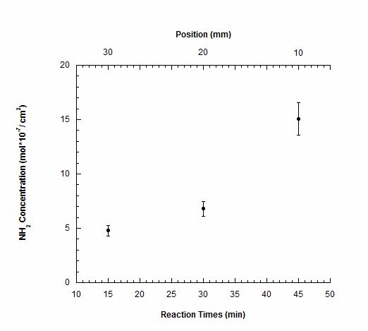
Figure 1. NH2 concentration as function of the reaction times and of the position along the PCL surface.
The PCL-NH2 substrates were treated with Rhodamine B Isothiocyanate to visualize the gradient by using CLSM. The fluorescence intensity decreased from 80 (a.u.) for the region treated for 45 minutes to 10 (a.u.) for the PCL zone. The region treated for 45 minutes showed a greater roughness, probably due to surface erosion of the material. (Fig. 2).
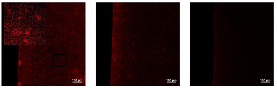
Figure 2. Representative images of PCL-NH2-Rhod surfaces functionalized after right) 45; middle) 30; and left) 15 minutes of treatment.
Static water contact angle decreased by about 15° whilst collagen density increased with increased NH2 concentration. The introduction of collagen improved the hydrophilicity of the surfaces. Microscopy imaging/analysis visualized the collagen distribution onto the substrates whilst FTIR confirmed collagen grafting and preservation of its helical structure.
Conclusion: A two-step functionalization procedure protein was developed. A continuous collagen gradient along the length of a PCL substrate was confirmed by morphological and physico-chemical analysis. Future studies will apply the methodology to FDM PCL-based scaffolds for the repair/regeneration of focal osteochondral defects.
The authors would like to thank FIRB MERIT project (GAE: P0000570) for providing financial support
References:
[1] Y. Zhu, Zheng, W. Mao, H.Y. Shi, C. Y. Gao, "In-depth study on aminolysis of poly(ɛ-caprolactone): Back to the fundamentals", Science China Chemistry. Vol. 55, Mar. 2012.
Keywords:
Tissue Engineering,
Surface modification,
Functionalization,
Polymeric material
Conference:
10th World Biomaterials Congress, Montréal, Canada, 17 May - 22 May, 2016.
Presentation Type:
Poster
Topic:
Regenerative medicine: biomaterials for control of tissue induction
Citation:
D'Amora
U,
D'Este
M,
Eglin
D,
Gloria
A,
De Santis
R,
Alini
M and
Ambrosio
L
(2016). Collagen density gradient on poly(epsilon-caprolactone) surface for the design of 3D printed osteochondral.
Front. Bioeng. Biotechnol.
Conference Abstract:
10th World Biomaterials Congress.
doi: 10.3389/conf.FBIOE.2016.01.02275
Copyright:
The abstracts in this collection have not been subject to any Frontiers peer review or checks, and are not endorsed by Frontiers.
They are made available through the Frontiers publishing platform as a service to conference organizers and presenters.
The copyright in the individual abstracts is owned by the author of each abstract or his/her employer unless otherwise stated.
Each abstract, as well as the collection of abstracts, are published under a Creative Commons CC-BY 4.0 (attribution) licence (https://creativecommons.org/licenses/by/4.0/) and may thus be reproduced, translated, adapted and be the subject of derivative works provided the authors and Frontiers are attributed.
For Frontiers’ terms and conditions please see https://www.frontiersin.org/legal/terms-and-conditions.
Received:
27 Mar 2016;
Published Online:
30 Mar 2016.