The use of microelectrodes to understand how neurons communicate has advanced our knowledge of how the brain functions. Metallic microelectrodes transduce ionic signals, associated with neuronal action potentials, into electronic signals through capacitive mechanisms[1]. Although successful, one major limitation is high impedance, which increases noise and distorts signal amplitude[2]. Conducting polymers such as poly(3,4-ethylenedioxythiophene (PEDOT) have shown promise as electrode coatings for metallic microelectrodes. The use of conducting polymer coatings increases the electrochemical surface area of an electrode, lowering impedance and therefore improving recording quality[3]. We aim to develop a low impedance macro-porous CP based sensor which is capable of detecting neuronal action potentials through changes in ionic concentrations (mainly Na+ and K+).
A custom fabricated microelectrode array (MEA) comprised of titanium/gold (40 nm/100 nm) electrical contacts and a SU8 photoresist insulating layer was constructed using conventional photolithographic protocols. SU8 defined the exposed electrode diameter at 25 µm. Non-porous and porous PEDOT (doped with polystyrene sulfonate (PSS)) structures were prepared through electrochemical polymerisation onto the gold microelectrodes. Porous PEDOT:PSS was prepared over a template of sacrificial polystyrene beads (ca. 230 nm) deposited on the microelectrodes. PEDOT structures were characterised for; electrical properties via electrochemical impedance spectroscopy (EIS) and cyclic voltammetry (CV), ionic sensitivity via flow through analysis of Na+ and K+ ions over a range of concentrations, structure via scanning electron microscopy (SEM) and biological compatibility of the MEA via the culture of primary hippocampal cells onto and around PEDOT structures.
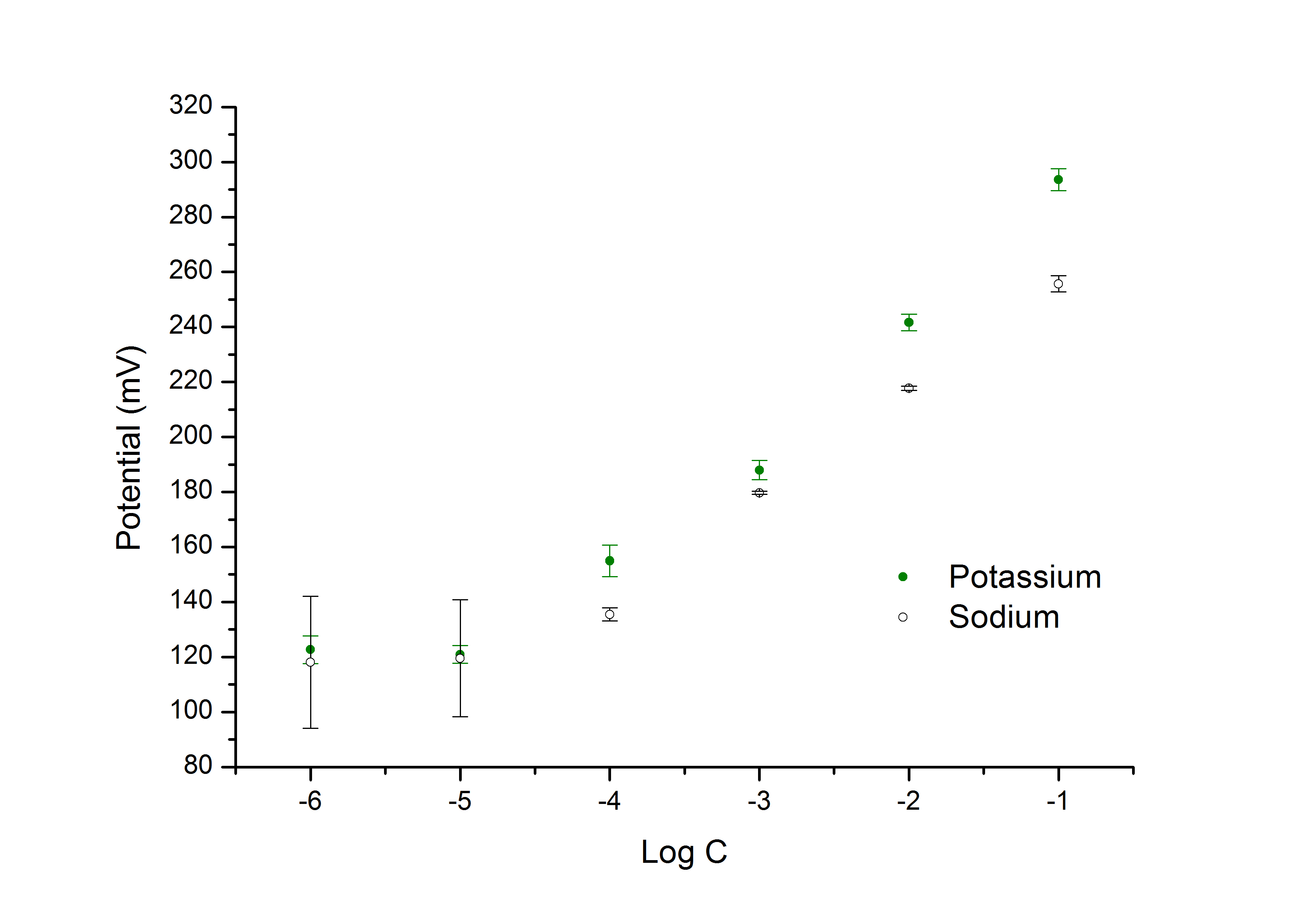
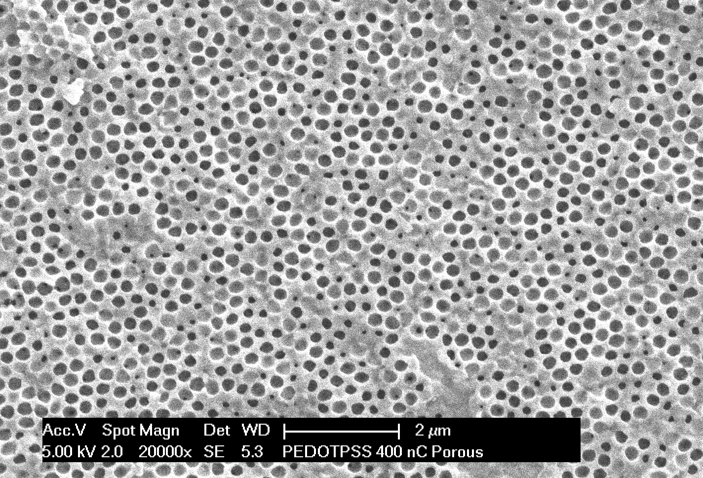
EIS revealed that PEDOT coatings successfully reduce electrode impedance from 395 kΩ, for bare gold, to 22 kΩ (at 1 kHz, which is characteristic of neuronal activity) and that the reduction in impedance was proportional to the amount of polymer deposited. CV studies showed that the polymer was electroactive and displayed reversible redox switching. PEDOT:PSS was able to detect changes in both Na+ and K+ ionic concentrations with near Nernstian responses (Figure 1). SEM images revealed that porous PEDOT:PSS structures were successfully synthesised onto the microelectrodes (Figure 2). Cultures of primary hippocampal cells onto the MEA showed that neurons were able to survive on and around PEDOT:PSS structures.
The impedance of gold microelectrodes was successfully reduced when coated with porous PEDOT:PSS. The PEDOT:PSS coatings were responsive to varying ionic concentrations of K+ and Na+ over 5 orders of magnitude (log -1 to log -5). Tests on 2 mM increment changes were also done, showing that it is possible for CPs to detect small ionic changes which occur during neuronal action potentials.

Figure 1: Open-circuit potential response of PEDOT:PSS (with 400 nC charge passed) to changes in potassium and sodium concentration

Figure 2: SEM image of macroporous PEDOT:PSS (with 400 nC of charge passed)
Neurological Foundation of New Zealand; The Royal Society of New Zealand - Marsden Fund; Auckland Medical Research Foundation
References:
[1] Cogan SF. Neural Stimulation and Recording Electrodes. Annual Review of Biomedical Engineering. 2008;10(1):275-309.
[2] Ludwig KA, Langhals NB, Joseph MD, Richardson-Burns SM, Hendricks JL, Kipke DR. Poly(3,4-ethylenedioxythiophene) (PEDOT) polymer coatings facilitate smaller neural recording electrodes. J Neural Eng. 2011;8(1):014001.
[3] Kip AL, Nicholas BL, Mike DJ, Sarah MR-B, Jeffrey LH, Daryl RK. Poly(3,4-ethylenedioxythiophene) (PEDOT) polymer coatings facilitate smaller neural recording electrodes. Journal of Neural Engineering. 2011;8(1):014001.