Introduction: Endogenous cell recruitment/homing is emerging as alternative to cell transplantation in regenerative medicine[1]. For homing, biomaterials are endowed with chemokines attracting cells capable of triggering a regeneration cascade from adjacent tissues. In this study we used a thermoresponsive hyaluronan hydrogel for delivering SDF-1 and CCL5, alone or in combination, to an osteochondral defect in rabbits.
Materials and Methods: The delivery system was prepared by grafting poly(N-isopropylacrylamide) to hyaluronan[2]. Temperature-induced gelation was verified via rheology with or without chemokines. In vitro release of SDF-1 and CCL5 from the hydrogel was performed for the experimental groups listed below up to 2 weeks using ELISA assays. Skeletally mature female New Zealand White rabbits of 4.1 ± 0.5 kg were enrolled in the in vivo study. A defect of 2.7 mm diameter and 4 mm depth was drilled in the central area of the medial trochlear ridge. The defect was either left empty (control) or filled with gel (H), gel + SDF-1 (S), gel + CCL5 (C), gel + SDF-1 + CCL5 (SC) for a total of 5 experimental groups and 6 animals per group and time interval. Chemokine concentration was 10 µg/ml (10+10 for the SC group). Cell density was quantified at 1 week counting DAPI stained cell nuclei in histological sections of the defect zone (n=6 per group). After 12 weeks the joints were evaluated macroscopically according to the ICRS score and histologically with the O'Driscoll score system.
Results: The addition of chemokines did not inhibit the temperature-induced gelation of the thermoresponsive hydrogel. In vitro release of SDF-1 plateaued at 1 week when 32% of the loaded chemokines was released. 50% CCL5 was released after 2 weeks. In vivo, the gel was not displaced from the defect. At 1week, significantly higher cell densities were found in the combination group SC vs H (p = 0.035) (fig 1) and all cytokine groups combined vs H (p ≤ 0.001). The interface between defect and native tissue for group CS at 1 week is illustrated in fig 2. Gross and histological scores were not significantly different between the chemokines containing group and the hydrogel group. However macroscopic score of the hydrogel containing groups was statistically superior (p=0.017) vs empty.
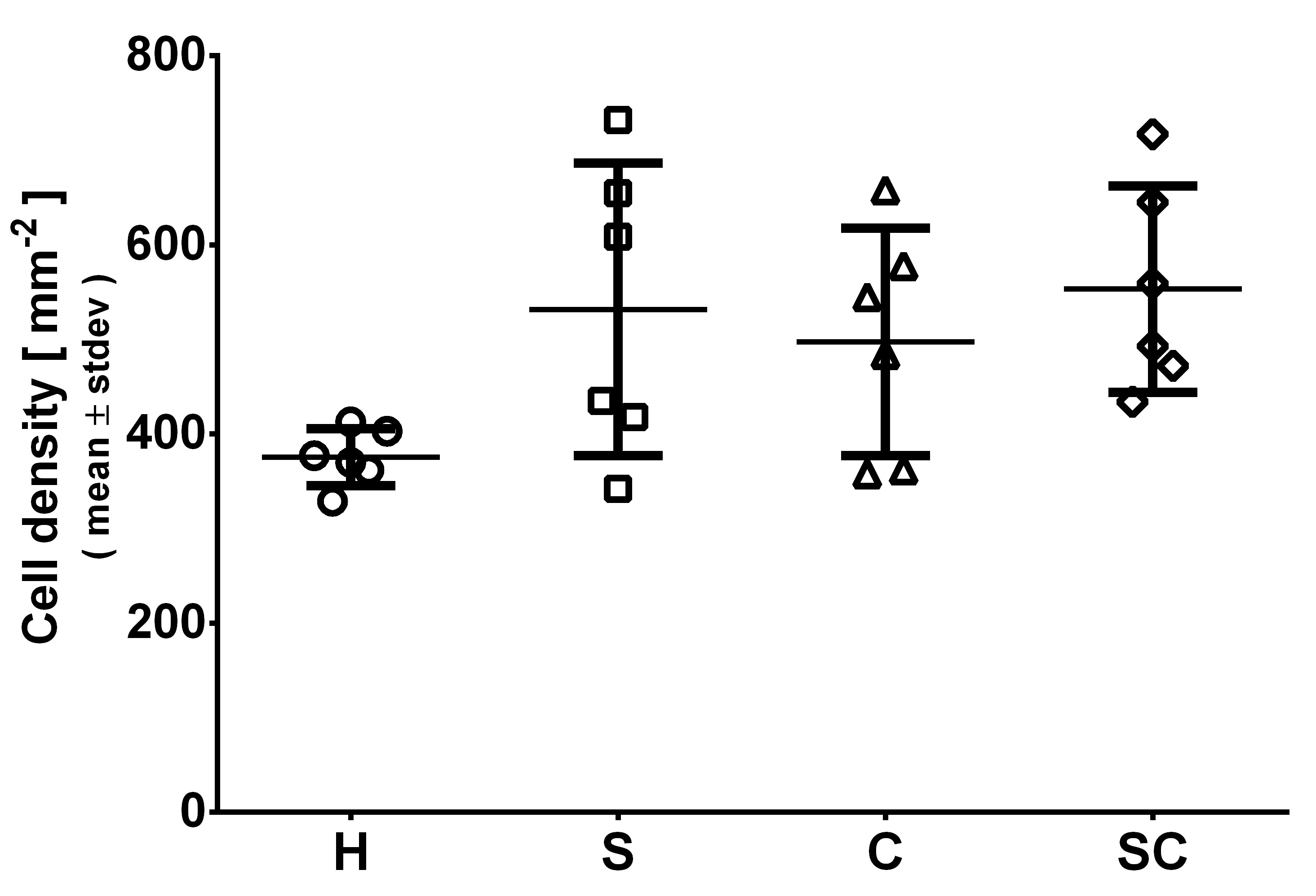
Figure 1: Box-plot illustrating the cell density within the subchondral defect area at 1 week. Data were tested for normal distribution and analysed by ANOVA. Statistical significance was set at p<0.05.
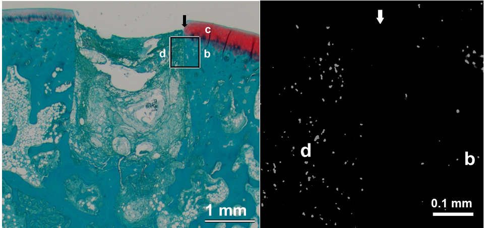
Fig. 2: Safranin O Fast Green staining (left)+DAPI (right, black and white-epifluorescence) image of the interface defect-native cartilage at 1 week for group CS.
Discussion: In presence of chemokines a higher cell density was observed in the defect suggesting the effective mobilisation of cells by the chemokines at 1 week. However, at 12 weeks, the repair of the hyaline cartilage tissue was not significantly different. It could be hypothesised that the cell mobilisation was not sufficient to enhance the repair process in the model used. Tissue repair accomplished was high for all groups; therefore the defect used in our model was subcritical.
Conclusion: SDF-1 and CCL5 delivered from thermoresponsive hyaluronan hydrogel attracted cells into an osteochondral defect in rabbit. In our model no significant difference in cartilage repair could be observed at 12 weeks. Optimization of pharmacokinetic and understanding of the cells mobilisation process is required to improve cartilage repair.
References:
[1] Ko IK, Lee SJ, Atala A, Yoo JJ.. Exp Mol Med. 2013;45:e57.
[2] M. D’Este, D. Eglin, M. Alini (2012) Carb Polym 90: 1378– 1385.