Introduction: Poly(lactic-co-glycolic acid) (PLGA), a copolymer of polylactic acid and polyglycolic acid, is one of the most commonly used biodegradable polymers in the field of tissue engineering. However, hydrophobicity, weak mechanical strength and poor bioactivity of PLGA are the critical issues of concern that often limit its applications[1]. The above mentioned limitations can be overcome by addition of suitable biocompatible reinforcement materials in the PLGA matrix to form biocomposite for bone tissue regeneration. Recently, carbon based biomaterials have attracted attention because of their nanoscale dimensions, large surface area and good acceptance by the biological environment[2]. Beyond the biological aspects, the mechanical properties of these biomaterials are also comparable to that of natural bone. Hence, it would be interesting to compare the behavior of 1-D and 2-D carbon nanomaterials reinforced PLGA nanocomposites. In the present work, the polymer matrix nanocomposites based on PLGA and two carbon allotropes, functionalized multi walled carbon nanotubes (CNT: 1-D, tubular) and graphene nanoplatelets (GNP: 2-D, sheets), were developed and studied to assess their potential use in bone tissue engineering.
Materials and Methods: PLGA films reinforced with 1 wt% of two different carbon nanomaterials, CNT and GNP, were prepared by solvent casting method. The developed nanocomposites were characterized using scanning electron microscopy (SEM), X-ray diffraction (XRD), fourier transform infrared spectroscopy (FTIR) and contact angle measurements. In addition, the effect of nanomaterials on in-vitro biological studies such as swelling, bioactivity, hemolysis and protein adsorption was evaluated. Further, these composites were tested for their mechanical strength and in-vitro biocompatibility. The SEM analysis, 3-(4,5-Dimethylthiazol-2-yl)-2,5-Diphenyltetrazolium Bromide (MTT) assay, Alkaline phosphatase (ALP) activity and Alizarin red S staining were performed to study the osteoblast (MG-63) cell proliferation and differentiation.
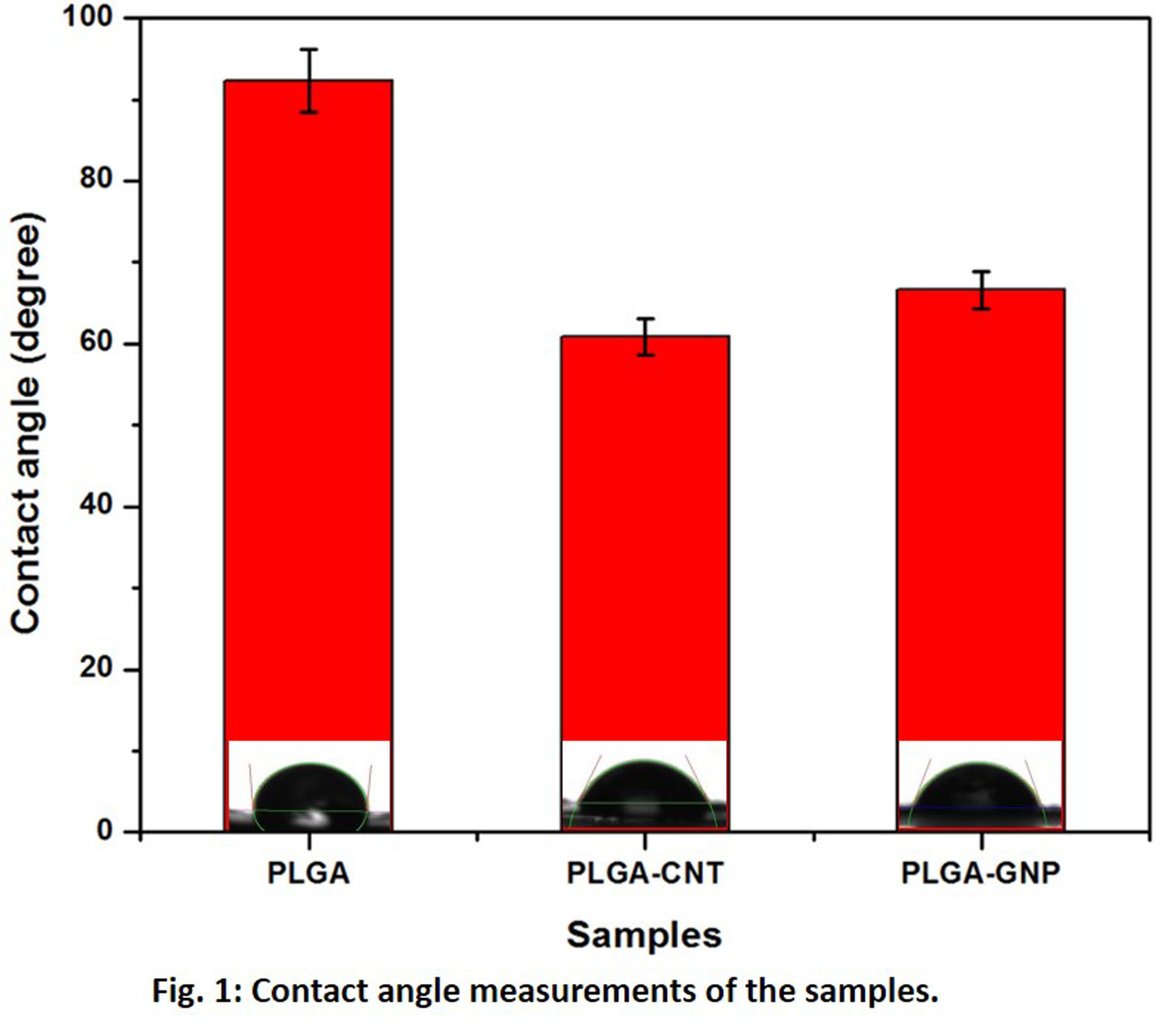
Results and Discussion: Both PLGA-CNT and PLGA-GNP nanocomposites exhibited improved hydrophilicity, bioactivity, protein adsorption and mechanical properties when compared to pure PLGA. The increased hydrophilicity (Fig. 1) with incorporation of CNT and GNP also promoted the cell biological behaviors (Fig. 2). The exceptional structural and electrical properties of the carbon nanomaterials led to enhanced osteoblast cell proliferation and differentiation. However, PLGA-GNP showed better results for mechanical properties and in-vitro biocompatibity when compared to PLGA-CNT. Higher cell viability (Fig. 2d), ALP activity and mineralization was observed in PLGA-GNP nanocomposites without the addition of any extra agents. This is due to the higher surface area provided by sheet like structure of GNP for more cells to adhere as compared to tubular CNT[3]. In contrast to CNT, the GNP were synthesized in pure form without any metallic impurities; hence, showed no toxicity to adherent cells.


Conclusion: All the results establish the practicability of using CNT and GNP as ideal reinforcement materials in PLGA for bone tissue engineering. However, the reinforcement of GNP resulted not only in improved mechanical properties but also showed better cell attachment and growth which is due to higher surface area when compared to CNT.
References:
[1] C. Lin, Y. Wang, Y. Lai, W. Yang, F. Jiao, H. Zhang, S. Ye and Q. Zhang, “Incorporation of carboxylation multiwalled carbon nanotubes into biodegradable poly(lactic-co-glycolic acid) for bone tissue engineering” Colloids and Surfaces B: Biointerfaces. Vol. 83, 2011.
[2] J.D. Núñez, A.M. Benito, R. Gonzalez, J. Aragon, R. Arenal and W.K. Maser, “Integration and bioactivity of hydroxyapatite grown on carbon nanotubes and graphene oxide” Carbon. Vol. 79, 2014.
[3] S. Duan, X. Yang, F. Mei, Y. Tang, X. Li, Y. Shi, J. Mao, H. Zhang and Q. Cai, “Enhanced osteogenic differentiation of mesenchymal stem cells on poly(L-lactide) nanofibrous scaffolds containing carbon nanomaterials” Journal of Biomedical Materials Research Part A. Vol. 103 2015.