Introduction: Hydroxyapatite [Ca10(PO4)6(OH)2; HA] is a well-known biomaterial constituent of biological hard tissues. Many bone tissue engineering scaffolds have been made as composites, by the introduction of HA nanoparticles within polymeric matrices. It is important to consider the possible impact of their composition on the processes linked to cell proliferation and activity, and to ensure the proportional amounts of each component do not impoverish the final composite properties. Hence, it would be of interest to evaluate the effect of different HA ratios of composite scaffolds on cell response[1].
Materials and Methods: The HA/GCPU porous scaffold was synthesized by glyceride of castor oil and isophorone diisocyanate (IPDI) with addition of HA powder (GCPU, HA20/GCPU and HA40/GCPU.). We focused on cell morphology, ultrastructural, and activity assays[2].
Results:

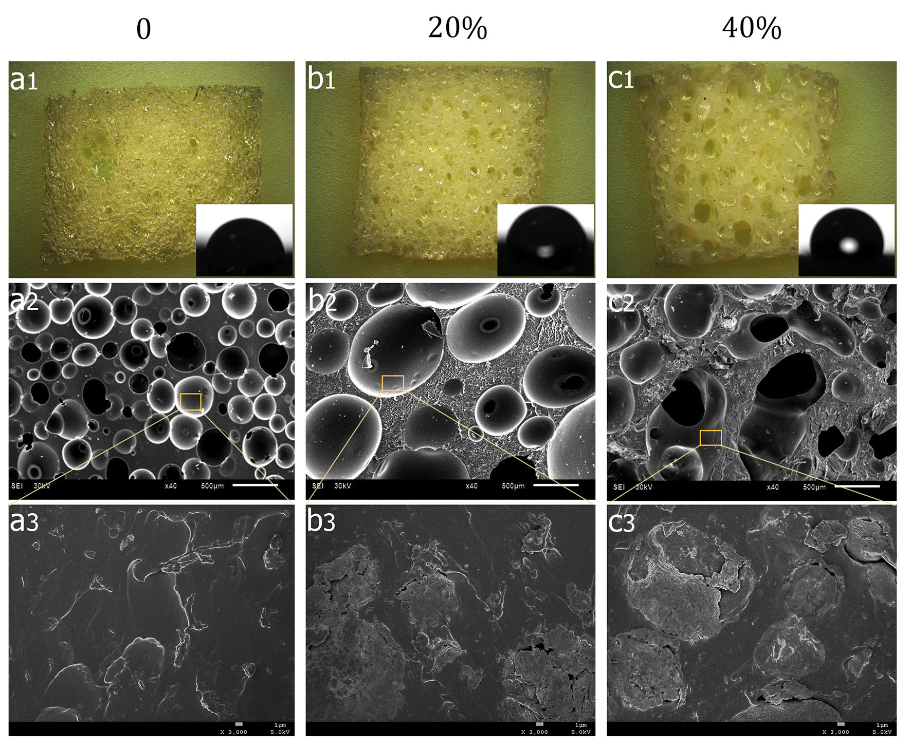
The HA was evenly distributed over the scaffold which exhibit good pore interconnectivity (Fig1. (a, b)). With the addition of HA, the contact angles became higher (about 69° to 91°). GCPU scaffolds showed a typical bubble structure with pore diameter from 100 μm to 400 μm(Fig1.(b1)). The macropore size of HA20 /GCPU scaffold was mainly ranging from 200 μm to 700μm, while the HA40/GCPU scaffold has an average pore size of 500 mm. GCPU scaffolds possessed a uniform surface with micro-wrinkles (Fig.a3) and globular HA deposits was increased with the addition of HA (Fig.(b3,c3)).

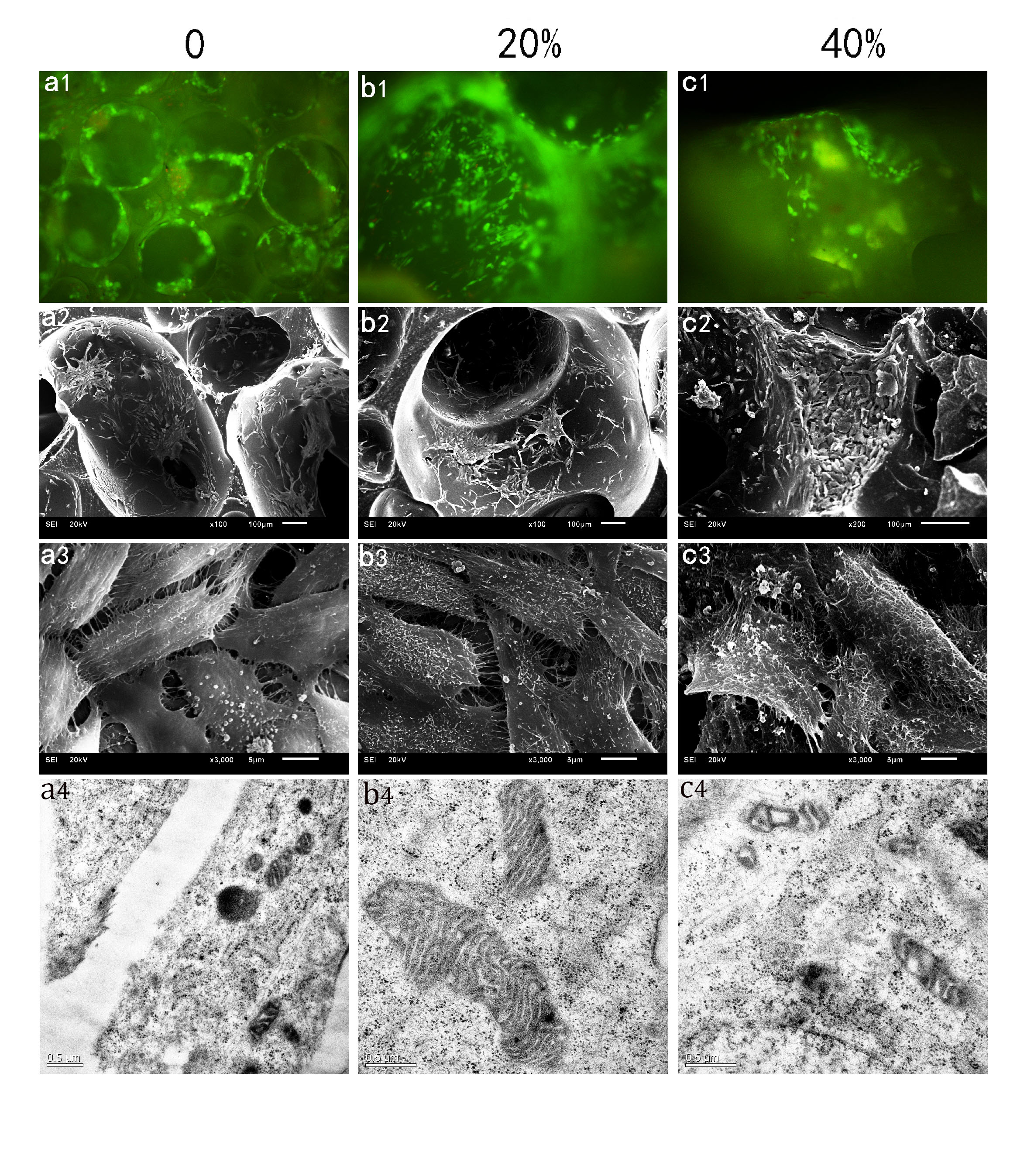
On GCPU smooth substrates a multilayer of cells was visible (Fig. 2.(a1)). The cells exhibited flattening on the substratum. Cells adhere to many parts on the HA20-GCPU scaffold surface (Fig. 2(b1)), especially displays multiple microvilli and numerous filopodia of cells on the surface (Fig. 2(b3)). Rough surfaces were observed due to the attachment of microparticles over the cell membrane on the HA40-GCPU specimen surfaces (Fig. 2(c2, c3)). Cells were rounded and had a ruffled morphology with lower cytoplasmic spreading (Fig. 2(C3)). The ultrastructural details of the cells on GCPU and HA20-GCPU scaffold surface have revealed no damaged cytoplasmic organelles, prominent structures like the nucleus, mitochondria, ribosomes and rough endoplasmic reticulum appeared to be well preserved(Fig.2 (a4, b4)). The cells on HA40-GCPU scaffold surface were found degeneration and vacuolization of the cytoplasm (Fig. 2(c4)).
Discussion: In our study enhanced cell activity was clearly manifested by MG-63 cells adhering to HA20-GCPU scaffold surfaces. On rougher surfaces cells might be able to attach directly to the surface. Moreover, modifying the wettability, closely related characteristics, protein adsorption and osteoblast proliferation and differentiation could be modulated[3]. Additionally, cell differentiation has been demonstrated by our observation of secretory-like vesicles[4] and a dominant presence of endoplasmatic reticulum is associated before with a high protein synthesis.
Conclusion: We used an osteogenic cell line as a model system and found that some structural surface parameters have a major impact on biological properties such as proliferation and viability. While the mechanism of HA-enhanced network formation in vitro is not clear, these findings have relevance to the development of biomaterials for bone tissue engineering.
Research Funds of China NSFC fund (No.31370971); Research Funds of Sichuan (No.2012FZ0125); Research Funds of Chengdu Project (No. 12DXYB145JH-05)
References:
[1] AbdulQader ST, Kannan TP, Rahman IA, Ismail H, Mahmood Z. Effect of different calcium phosphate scaffold ratios on odontogenic differentiation of human dental pulp cells. Materials science & engineering C, Materials for biological applications 2015;49:225-33
[2] Du JJ, Zuo Y, Zou Q, Sun B, Zhou MB, Li LM, et al. Preparation andin vitroevaluation of polyurethane composite scaffolds based on glycerol esterified castor oil and hydroxyapatite. Materials Research Innovations 2014;18:160-8
[3] Montesi M, Panseri S, Iafisco M, Adamiano A, Tampieri A. Effect of hydroxyapatite nanocrystals functionalized with lactoferrin in osteogenic differentiation of mesenchymal stem cells. Journal of biomedical materials research Part A 2015;103:224-34
[4] Padial-Molina M, Galindo-Moreno P, Fernandez-Barbero JE, O'Valle F, Jodar-Reyes AB, Ortega-Vinuesa JL, et al. Role of wettability and nanoroughness on interactions between osteoblast and modified silicon surfaces. Acta biomaterialia 2011;7:771-8