Introduction: Studies have shown that the surface physicochemical properties of biomaterials had profound influence on cell behaviors, but the interactions between material surfaces and cells have not been fully understood[1]. Although TiO2 nanotube arrays prepared by anodization are beneficial to cell adhesion, proliferation, etc. due to their unique structures and properties[2], the influence of TiO2 nanotube arrays with different diameters on cell behaviors is still under dispute[3],[4]. Previous studies focused on the effects of TiO2 nanotube arrays with uniform and small diameters (≤100nm) on cell behaviors. In this work, we prepared large diameter (100-500 nm) TiO2 nanotube arrays and coexisting multi-size TiO2 nanotube arrays by anodization, and investigated their influences on cell behaviors.
Materials and Methods: Gradient voltage- increasing anodization was used to prepare large diameter TiO2 nanotube arrays, and coexisting multi-size TiO2 nanotube arrays were fabricated by repeated anodization. Titanium foil served as anode, and stainless steel was used as cathode. The electrolyte consisted of NH4F, H2O in ethylene glycol. Highly ordered TiO2 nanotube arrays were fabricated at voltages of 60V-150V. The titanium foil was divided into four regions, and each region was anodized at 15V, 40V, 80V and 120V to prepare the coexisting multi-size TiO2 nanotube arrays. The morphologies of samples were characterized by SEM. MC3T3-E1 cells were cultured on the surface of samples mentioned above, and the cell behaviors were observed by SEM, fluorescent staining, MTT, ALP activity, etc.
Results and Discussion: Highly ordered TiO2 nanotube arrays with diameter of 150nm, 260nm, 360nm, 470nm were prepared at voltages of 60V, 90V, 120V, 150V, respectively (Fig. 1Ⅰ). For coexisting multi-size TiO2 nanotube arrays (Fig.1Ⅱ), the TiO2 nanotubes were regular at the boundaries between two different regions, and no obvious difference in height between two regions existed. The results of ALP test showed that the highest ALP activity appeared on the 150 nm samples, and the activity decreased gradually as tube diameter increased (See Fig. 2Ⅰ). The heat maps (Fig. 2Ⅱ) showed the cell density distribution after incubation of 2h, 6h, 12h, and 24h on coexisting four-diameter TiO2 nanotube arrays, respectively. After 2h incubation, the highest cell density appeared on 60nm diameter, and the lowest area was 350nm. At 24h, the cell density on 60nm and 150nm was obviously higher than that on 250nm and 350nm. We believed that the small diameter nanotubes provided more anchor points for proteins attachment, which leaded to more cells adhering on small diameter nanotubes.

Fig. 1(Ⅰ) SEM surface morphologies of TiO2 nanotube arrays anodized at 60 V, 90 V, 120 V, 150 V, 180 V, respectively, associated with diameters range from 150 nmto 570 nm; (Ⅱ) The boundary morphologies between two different diameters of coexisting four-diameter TiO2 nanotube arrays: (a) 60 nm and 150 nm; (b) 150 nm and 250 nm, (c) 60 nm and 350 nm, (d) 250 nm and 350 nm. (All insets show partial enlargement of boundary morphologies).
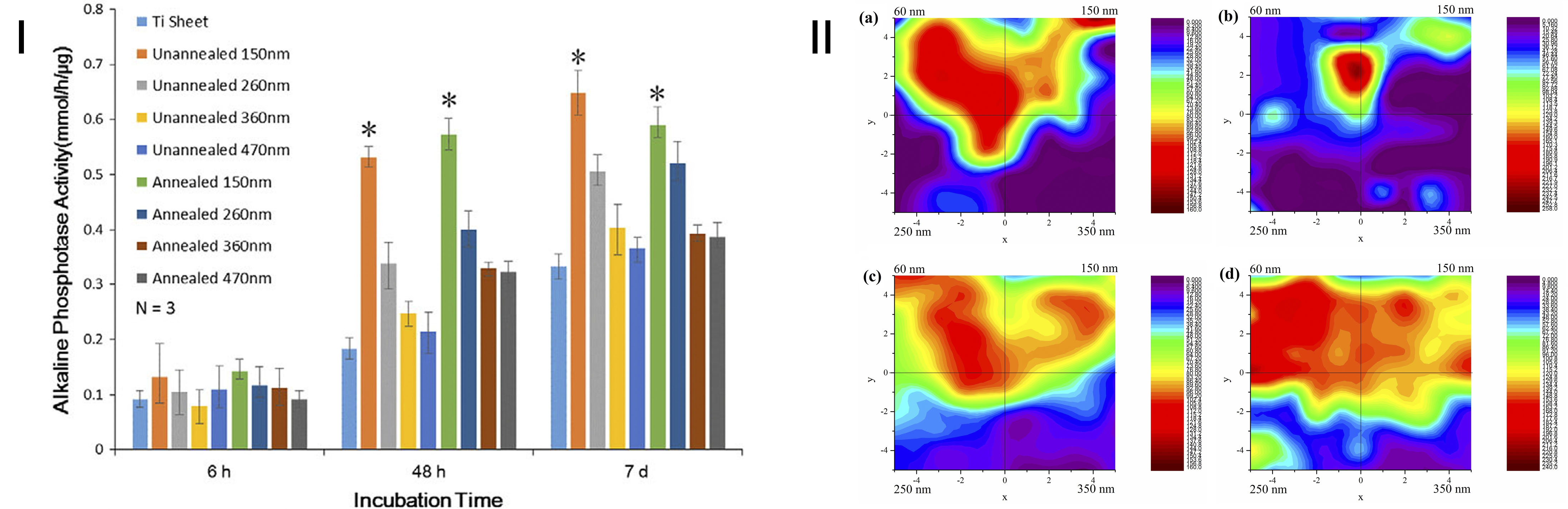
Fig. 2 (Ⅰ) The results of ALP on the surfaces of annealed and unannealed TiO2 nanotube arrays. The bar graphs showmean±SD. * indicates a significant difference between 150 nmsample and all other samples in the same time slot (p b 0.05); (Ⅱ) Heat map of cell density distribution on coexisting four-diameter TiO2 nanotube arrays after incubation of (a) 2 h; (b) 6 h; (c)12 h; (d) 24h (Red shows the highest cell density, Purple shows the lowest cell density).
Conclusions: In this work, large diameter (150-500nm) TiO2 nanotube arrays and coexisting four-diameter TiO2 nanotube arrays were fabricated by modified anodization. For large diameter TiO2 nanotube arrays, cell spread fully on 470 nm diameter, and the highest ALP activity appeared on 150 nm diameter. For coexisting four-diameter TiO2 nanotube arrays, smaller diameter offered the better triggering structure for cells adhesion than the larger diameter nanotubes under the coexistence situation of different diameters.
National Natural Science Foundation of China (No. 51401126, No. 51271117) and Shanghai Committee of Science and Technology, China (No. 14441901800)
References:
[1] Ventre M, Cause F, Netti P A. Determinants of cell-material crosstalk at the interface: towards engineering of cell instructive materials. J. R. Soc. Interface. 2012, 9: 2017-2032.
[2] Park J, Bauer S, Schlegel K A, Neukam F W, Von der Mark K, Schmuki P. TiO2 nanotube surfaces: 15 nm-An optimal length scale of surface topography for cell adhesion and differentiation. Small 2009, 5(6): 666-671.
[3] Oh S, Brammer K S, Julie Li Y S, Teng D Y, Engler A J, Chien S, Jin S. Stem cell fate dictated solely by altered nanotube dimension. Proc. Natl. Acad. Sci. U. S. A. 2009, 106 (7): 2130-2135.
[4] Park J, Bauer S, Mark V D, Schmuki P. Nanosize and vitality: TiO2 nanotube diameter directs cell fate. Nano Lett. 2007, 7 (6):1686-1691.