Effect of poly-L-co-D,L-lactide scaffolds combined with simvastatin and mesenchymal cells on calvarial bone defects and ectopic subcutaneous bone formation
-
1
Pontifical Catholic University of São Paulo, Medical student, Faculty of Medical Sciences and Health, Brazil
-
2
Pontifical Catholic University of São Paulo, Laboratory of Biomaterials, Faculty of Medical Sciences and Health, Brazil
-
3
Pontifical Catholic University of São Paulo, Morphology and Pathology Department, Faculty of Medical Sciences and Health, Brazil
-
4
Pontifical Catholic University of São Paulo, Physiology Department, Faculty of Medical Sciences and Health, Brazil
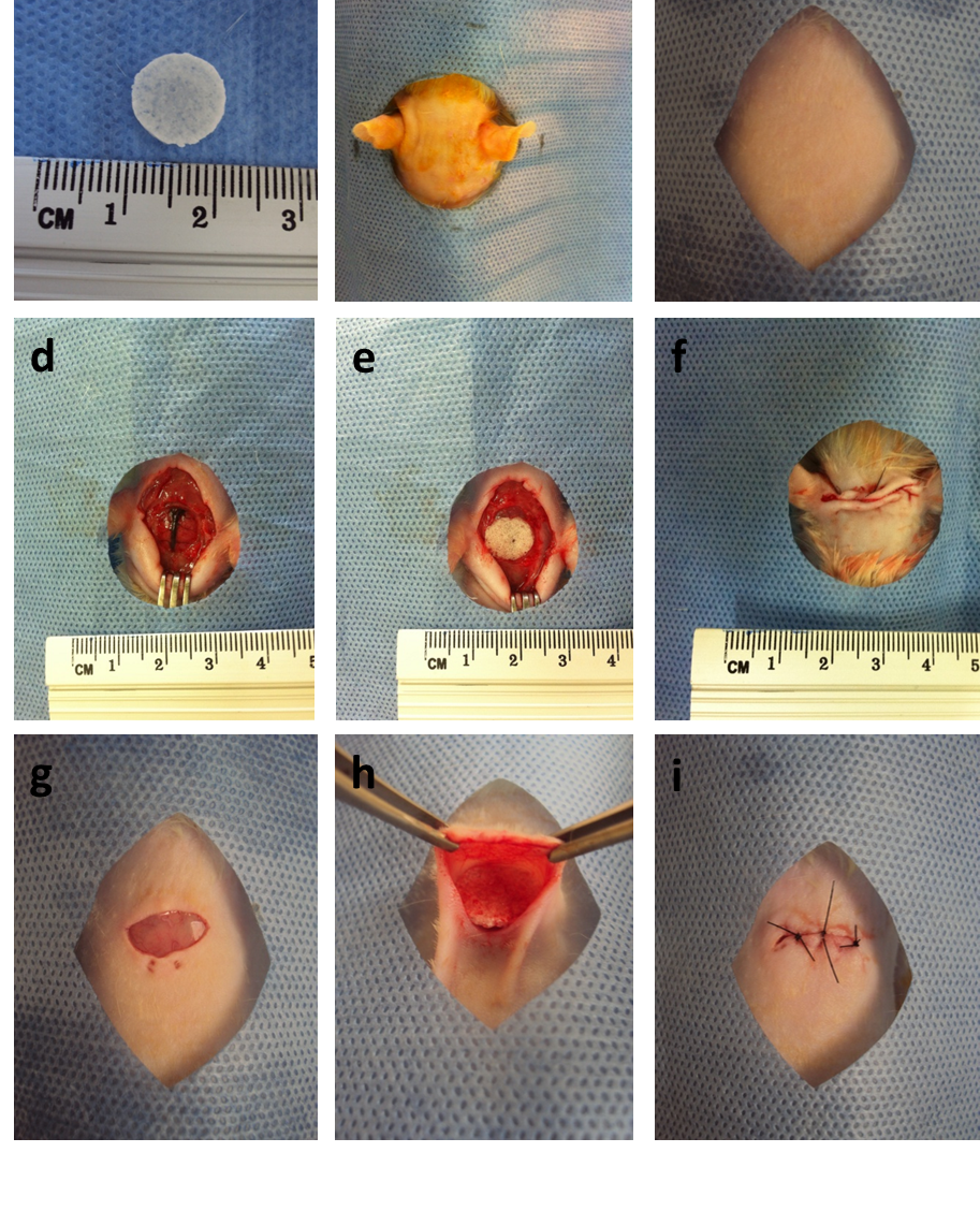
Tissue bioengineering has become popular as a method of repair of damaged tissues, and allows for both an aesthetic and functional result[1]. One of the biomaterials used in the treatment of bone defects are the hydrolytically degradable polymers, specifically the poly-L-co-D,L-lactide (PLDLA)[2]. PLDLA is considered an attractive material for drug delivery systems, such as for simvastatin release. Several studies show that statins have the capacity of stimulating bone formation by increasing expression of bone morphogenetic protein-2 (BMP-2)[3]. Mesenchymal stem cells (MSC) are also involved in the process of bone growth, since they have potential to differentiate into osteoblasts and osteocytes[4]. Based on these concepts, this study analyzed the effect of PLDLA scaffolds with simvastatin and MSC in regards to their capacity to promote bone formation in both calvarial defects and the subcutaneous tissues of Wistar rats[5]. The 98 animals used for the study were divided according to the area of scaffold implantation, time of euthanasia and treatment applied. The implant areas were collected post-euthanasia and submitted for histologic analysis. New bone formation was detected in the animals who received calvarial implants, with statistical significance. In comparison to negative control, new bone tissue was especially observed in the groups receiving simvastatin and MSC (p < 0.01). No ectopic bone growth was seen in the subcutaneous tissue. The bio-affinity of the polymeric membrane and the osteogenic character of simvastatin and MSC were confirmed in this study.
References:
[1] Miguel FB, Cardoso AKMV, Barbosa AA, et al. Morphological assessment of the behavior of three-dimensional anionic collagen matrices in bone regeneration in rats. J Biomed Mater Res 2006; 78(2):334-339.
[2] Coimbra MER, Elias CN, Coelho PG. In vitro degradation of poly-l-d-lactic acid (PLDLA) pellets and powder used as synthetic alloplasts for bone grafting. J Mater Sci Mater Med 2008; 19(10): 3227-3234.
[3] Anbinder AL, Junqueira JC, Mancini MNG, et al. Influence of simvastatin on bone regeneration of tibial defects and blood cholesterol level in rats. Braz Dent J 2006; 17(4): 267-273.
[4] Heino TJ, Hentunen TA. Differentiation of osteoblasts and osteocytes from mesenchymal stem cells. Curr Stem Cell Res Ther 2008; 3(2): 131-145.
[5] Houshmand B, Hassanizade R, Eslami B, et al. Simvastatin and lovastatin induce ectopic bone formation in rat subcutaneous tissue. J Periodont Implant Dent 2010; 2(1): 12-16.
Keywords:
Bone Regeneration,
stem cell,
Drug delivery,
biomaterial
Conference:
10th World Biomaterials Congress, Montréal, Canada, 17 May - 22 May, 2016.
Presentation Type:
Poster
Topic:
Biomaterials for therapeutic delivery
Citation:
Edelmuth
SV,
Ramos
KS,
Aoki
KS,
Gaiotto
KK,
Más
BA,
Barbo
ML,
Duek
EA and
Oliveira
NM
(2016). Effect of poly-L-co-D,L-lactide scaffolds combined with simvastatin and mesenchymal cells on calvarial bone defects and ectopic subcutaneous bone formation.
Front. Bioeng. Biotechnol.
Conference Abstract:
10th World Biomaterials Congress.
doi: 10.3389/conf.FBIOE.2016.01.01030
Copyright:
The abstracts in this collection have not been subject to any Frontiers peer review or checks, and are not endorsed by Frontiers.
They are made available through the Frontiers publishing platform as a service to conference organizers and presenters.
The copyright in the individual abstracts is owned by the author of each abstract or his/her employer unless otherwise stated.
Each abstract, as well as the collection of abstracts, are published under a Creative Commons CC-BY 4.0 (attribution) licence (https://creativecommons.org/licenses/by/4.0/) and may thus be reproduced, translated, adapted and be the subject of derivative works provided the authors and Frontiers are attributed.
For Frontiers’ terms and conditions please see https://www.frontiersin.org/legal/terms-and-conditions.
Received:
27 Mar 2016;
Published Online:
30 Mar 2016.
*
Correspondence:
Dr. Stephanie V Edelmuth, Pontifical Catholic University of São Paulo, Medical student, Faculty of Medical Sciences and Health, Sorocaba, Brazil, bijuleao@hotmail.com
Dr. Kennedy S Ramos, Pontifical Catholic University of São Paulo, Medical student, Faculty of Medical Sciences and Health, Sorocaba, Brazil, kennedy.ramos@me.com
Dr. Khatharine S Aoki, Pontifical Catholic University of São Paulo, Medical student, Faculty of Medical Sciences and Health, Sorocaba, Brazil, katy.aoki@hotmail.com
Dr. Karlla K Gaiotto, Pontifical Catholic University of São Paulo, Medical student, Faculty of Medical Sciences and Health, Sorocaba, Brazil, karllagaiotto@hotmail.com
Dr. Bruna A Más, Pontifical Catholic University of São Paulo, Laboratory of Biomaterials, Faculty of Medical Sciences and Health, Sorocaba, Brazil, masbruna@yahoo.com.br
Dr. Mona L Barbo, Pontifical Catholic University of São Paulo, Morphology and Pathology Department, Faculty of Medical Sciences and Health, Sorocaba, Brazil, lourdesperis@uol.com.br
Dr. Eliana A Duek, Pontifical Catholic University of São Paulo, Physiology Department, Faculty of Medical Sciences and Health, Sorocaba, Brazil, eliduek@fem.unicamp.br
Dr. Newton M Oliveira, Pontifical Catholic University of São Paulo, Morphology and Pathology Department, Faculty of Medical Sciences and Health, Sorocaba, Brazil, newtonmo@hotmail.com