Three-dimensional (3D) multicellular aggregates are plausible to characterize and evaluate phenotype of malignant tumor in biologically relevant configuration. Indeed, solid tumors grow in spatially heterogeneous microenvironments (i.e., oxygen, nutrients, or mechanical stresses). Three-dimensional tumorspheres (TS) can reflect this diffusion-limited heterogeneity of solid tumors in vivo, compared to 2D monolayers. Although numerous approaches for tumorsphere (TS) formation have developed in cancer research to take into account the microenvironments and architectures, most methods provide a floating non-adherent multicellular cell aggregates. However, floating TSs is limited to present the invasive ability toward the surrounding microenvironment.
Here, we proposed 3D tumorsphere-based invasion assay platform consisting of two different hydrogels: a bio-inert (non-adherent) hydrogel for spontaneous formation of TS and a subsequent bio-active ECM (cell-interactive) hydrogel for evaluation of TS invasion. Using a small subpopulation of cells within malignant brain tumor, the glioma cancer stem cells (GSCs), we observed the formation of human GSC-TS and subsequent invasion of TS.
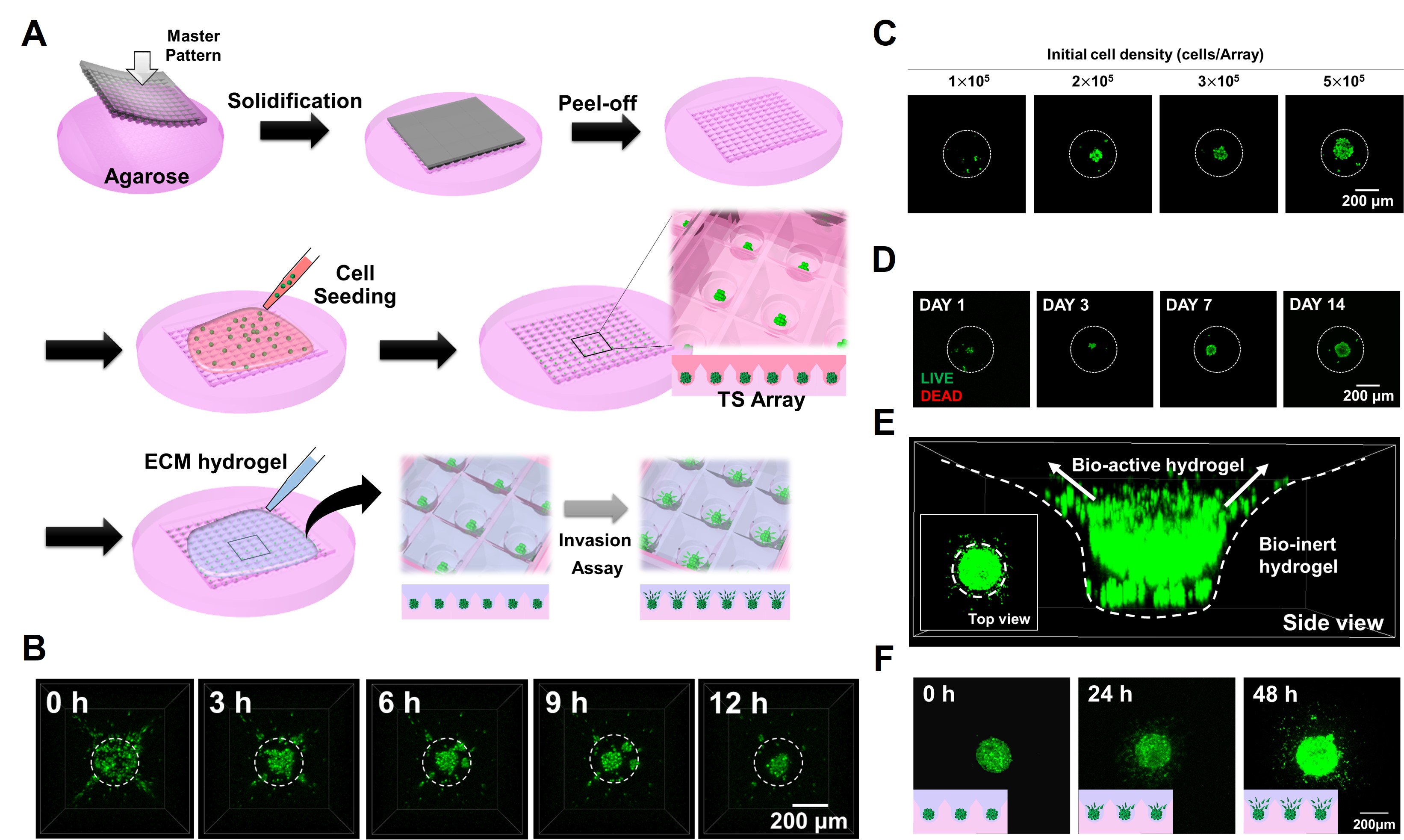
The invasion assay platform was fabricated by replica molding, as described in Figure 1A. First of all, a bio-inert agarose hydrogel was fabricated from the master pattern. GSCs suspensions isolated from specimen of patients were seeded on the agarose hydrogel platform. The isolated cells within a platform spontaneously formed sphere within a day (Figure 1B).
Within the platform, the size of TS was controlled by manipulating the initial seeding density as shown in Figure 1C. As tumorspheres grow, due to diffusion-limited heterogeneity in oxygen and nutrients, hypoxic core is spontaneously formed (Figure 1D). We further investigated that the hypoxia within GSC TS was positively correlated with tumor invasiveness. Moreover, to study ECM-TS interactions, we introduced a biologically reactive ECM hydrogel in the agarose hydrogel-based TS array platform (Figure 1A). Since the initially formed TS can only interact with upper ECM hydrogel, not lower bio-inert hydrogel, the invasive cancer cells directionally invade into the ECM hydrogel; the direction of cancer cell invasion can be controlled (Figure 1E). We thus observed the 3D cancer cell invasion into two-dimensional window. Bi-layered TS array platform provides easier visual inspection, enabling quantitative analysis on invasiveness of cancer cells in a 3D system (Figure 1F).
Collectively, Bi-layered 3D tumorsphere array platform enable us to observe biomimetic configurations and interactions between TS and the surrounding ECM microenvironment, consequently providing quantitative analysis of the TS invasion in high-throughput manner. Therefore, our platform can be applicable as a drug evaluation tool in preclinical screening phases of an anti-cancer drug discovery by investigating the invasive ability of malignant brain tumors, as well as other systematic cancers.
This research was supported from the National Research Foundation of Korea(NRF) (grant number : NRF-2015H1A2A1030560 and NRF-2014R1A1A1004985) and by a grant of the Korea Health Technology R&D Project through the Korea Health Industry Development Institute (KHIDI), funded by the Ministry of Health & Welfare, Republic of Korea (grant number : HI14C0042).
References:
[1] J. Dahlmann, G. Kensah, H. Kempf, D. Skvorc, A. Gawol, D. A. Elliott, G. Drager, R. Zweigerdt, U. Martin, and I. Gruh, “The use of agarose microwells for scalable embryoid body formation and car-diac differentiation of human and murine pluripotent stem cells,” Biomaterials, 34, 2013
[2] K. M. Charoen, B. Fallica, Y. L. Colson, M. H. Zaman, M. W. Grinstaff, “Embedded multicellular spheroids as a biomimetic 3D cancer model for evaluating drug and drug-device combinations,” Biomaterials, 35, 2014
[3] M. Lee, K. Yang, Y. H. Hwang, Y. Byun, D. Y. Lee, S, -W. Cho, and H. Lee, “Spheroform: Thera-peutic spheroid-forming nanotextured surfaces inspired by desert beetle Physosterna cribripes,” Ad-vanced Healthcare Materials, 4, 2015