Introduction: Time-of-flight secondary ion mass spectrometry (TOF-SIMS) is an established, highly sensitive analytical technique for mass spectrometry (MS) imaging applications with a lateral resolution below 100 nm. Monitoring the uptake of nanoparticles, drugs or other chemicals into cells are only a few examples for the application of this label-free chemical analysis technique. Chemical information is obtained by bombarding the surface with a focused primary ion beam and analysing the generated secondary ions in a TOF mass analyzer.
However in complex biological samples identification of unknown compounds can be hampered by mass interferences and a high number of possible candidates for a single mass peak.
In order to overcome these limitations, the 3D nanoSIMS project[1] is developing a revolutionary new SIMS instrument that combines the high lateral resolution and speed associated with TOF-SIMS with the high mass resolution and high mass accuracy of an orbital trapping mass analyser. The instrument is equipped with a newly developed gas cluster ion beam column allowing a lateral resolution down to the micron level. First results obtained from different biomaterials are presented here.
Materials and Methods: Amiodarone-dosed NR8383 cells and a native coronal mouse brain section were analysed using either a 20 keV argon gas or a bismuth cluster primary ion beam. For MS of the generated secondary ions a new hybrid SIMS instrument was utilized, featuring a hybrid mass analyser that combines a fast TOF analyser (TOFSIMS.5, ION-TOF GmbH, Münster, Germany) with an orbital trapping analyser (QExactiveTM HF[2], Thermo Scientific, Bremen, Germany).
Results and Discussion:
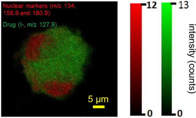
Fig. 1: TOF-SIMS image showing the uptake of the drug amiodarone into a single macrophage[3].
The high lateral resolution TOF-SIMS image in Fig. 1 shows the sub-cellular distribution of iodine, a moiety of the drug amiodarone (green) with respect to the nucleus of the cell (red).
First gas cluster OrbitrapTM SIMS images from mouse brain sections demonstrate the simultaneous localization and identification of various compounds. On tissue, the high mass resolution (FWHM of 240 000 at m/z 200) was used to identify lipids and metabolites with sub-ppm mass accuracy. Identification of numerous lipid signals at the single cell level could be easily performed using exact mass measurement and comparison with databases. Additionally ions were selectively fragmented by tandem MS (MS/MS) in order to confirm chemical structure or help on assignment of unknown signals.
Conclusion: With this unique instrument numerous applications in the field of biomaterials can be studied, including interactions between tissue and biomaterials and process control in fabrication. Laterally resolved, label-free, and non-targeted imaging of biomaterials at micron resolution is now possible.
References:
[1] The 3D nanoSIMS project, http://www.npl.co.uk/news/3d-nanosims-label-free-molecular-imaging
[2] Scheltema, et al. Mol Cell Proteomics (2014).
[3] Passarelli, et al. Anal. Chem. (2015).