Introduction: Bioactive hydrogels show great promise in tissue engineering and clinical applications. Collagen-based hydrogels, such as gelatin methacrylate (GelMA), contain amino acid sequences that mimic portions of native extracellular matrix and thus display excellent biocompatibility. As a photocrosslinkable polymer, GelMA also enables the study of cells in three dimensional culture[1]. Nanodiamonds (NDs), carbon-based nanoparticles displaying low cytotoxicity, have been used to bind small molecules, proteins, and DNA for drug delivery or gene therapy purposes[2]. Combining nanodiamonds with gelatin methacrylate to form a photocrosslinkable nanocomposite creates a flexible tissue engineering and drug delivery platform enabling investigation of ND/drug complexes in 3D culture. The goal of this study was to create GelMA/ND nanocomposites and characterize their structural properties and biocompatibility in vitro with human adipose stem cells (hASC) for regenerative medicine applications.
Materials and Methods: Gelatin methacrylate was synthesized by addition of methacrylic anhydride to a solution of 10% gelatin from porcine skin, which was then dialyzed for 5 days with 10 water changes, then frozen and lyophilized. Nanodiamonds at concentrations of 0.5, 1, and 2 mg/mL were dispersed by sonication in GelMA solutions for 30 minutes. Addition of .5% photoinitiator (Irgacure 2959) followed by 30 minutes of UV exposure resulted in nanocomposite gelation. Structural and porosity analysis of the resulting gels was performed using scanning electron microscopy and ImageJ. Mechanical studies to determine elastic modulus were performed using dynamic mechanical analysis. Swelling ratio was determined at 1, 3, 5, 24, and 48 hours. For cell studies, hASCs were seeded on the resulting gels for metabolic activity quantification by MTT, F-actin staining with phalloidin, and nucleus staining with DAPI. Cell area analysis was performed after 24 hours.
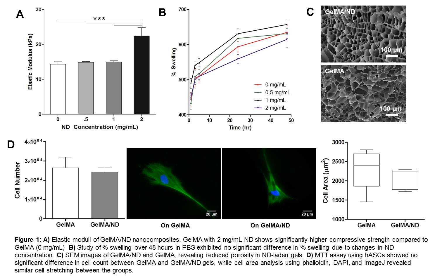
Results and Discussion: Mechanical analysis showed similar elastic moduli between GelMA, 0.5, and 1 mg/mL gels, while 2 mg/mL ND gels had significantly higher elastic moduli (Fig 1) while 4 mg/mL gels showed significant reduction in mechanical properties (not shown), meaning NDs show some potential for modulating GelMA mechanical properties. ImageJ Analysis of SEM samples (Fig 1) showed an average porosity of 44 ± 2% for GelMA/ND gels and 59 ± 6% for GelMA controls. Determination of the swelling ratio of 0.5, 1, and 2 mg/mL gels suggests that NDs do not significantly the diffusive properties of the GelMA. The MTT assay revealed no significant difference in cytocompatibility between GelMA and GelMA with 1 mg/mL ND, emphasizing the biocompatibility of NDs with the hASCs. Cell area analysis showed similar cell stretching between the GelMA and 1 mg/mL ND groups, further confirming cytocompatibility. These results suggest that NDs have the potential to manipulate mechanical properties with changing concentration and can be used as a drug delivery vehicle.
Conclusion: Nanocomposites comprised of GelMA and NDs were successfully synthesized and characterized for mechanical properties, structure, and biocompatibility. This system can thus be used as a tissue engineering platform to study cell-nanomaterial, cell-drug, and cell-hydrogel interactions in a 2D and 3D environment.
Arghya Paul likes to acknowledge the Institutional Development Award (IDeA) from the National Institute of General Medical Sciences, National Institutes of Health (NIH), under Award Number P20GM103638.
References:
[1] Mochalin VN, Shenderova O, Ho D, Gogotsi Y (2011). The properties and applications of nanodiamonds. Nat Nanotechnol. 7(1):11-23.
[2] Nichol JW, Koshy ST, Bae H, Hwang CM, Yamanlar S, Khademhosseini A (2010). Cell-laden microengineered gelatin methacrylate hydrogels. Biomaterials. (21):5536-44.