Introduction: Calcium phosphate particles intended as bone fillers and drug delivery agents are gaining interest worldwide. Size of the particles becomes an important factor when they are intended to act as site specific delivery agents, for therapeutic purposes. Cellular uptake, velocity of drug carrier in physiological pathways and specificity towards binding of therapeutic molecules are some key parameters that are greatly influenced by particle size[1]. The aim of the current study was to produce calcium phosphate particles through a modified electrospraying process[2]; characterize them physicochemically, evaluate their cytocompatibility and to analyse the efficacy of the particles to act as drug delivery systems.
Materials and Methods: Calcium nitrate tetrahydrate (CNt) and triethyl phosphite (TEP) were used as the calcium and phosphorous precursors with an aqueous solvent. Sols with the required Ca/P ratio (1.67 & 1.61) were prepared by adding CNt solution drop wise to the hydrolysed TEP solution. The obtained sols were aged and subsequently blended with a polyvinylalcohol solution. The solution was loaded into a 10ml syringe fitted with an 18G blunted needle. The tip to collector distance, flow rate and voltage were optimised for electrospraying. The as sprayed particles were dried overnight and then subjected to a controlled heating upto 500˚C. The particles obtained on heating 1day and 5days aged sols of 1.67 and 1.61 Ca/P ratio will be referred to as CaP167-1d, CaP167-5d, CaP161-1d and CaP161-5d respectively. Phase confirmation was done through X-ray diffraction (XRD). Stability of phases was tested by heating upto 900˚C. The particles size and morphology were analysed using transmission electron microscopy (TEM). In vitro biological evaluation of the particles were performed using L-929 and HOS cell lines. The drug delivering capacities of the particles were assessed using tetracycline as model drug.
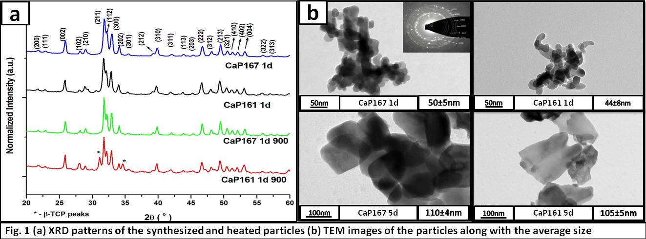
Results and Discussion: Figure 1 shows the results of XRD and TEM analysis. XRD patterns were matched with that of hydroxyapatite (JCPDS file no. 9-432). At 500˚C the patterns of all the particles are similar whereas on heating to 900˚C, CaP167-1d and 5d are the same while CaP161-1d and 5d develop peaks of β-tricalcium phosphate. From TEM imaging it is evident that increasing the aging time of sols has lead to an increase in the electrosprayed particle size.
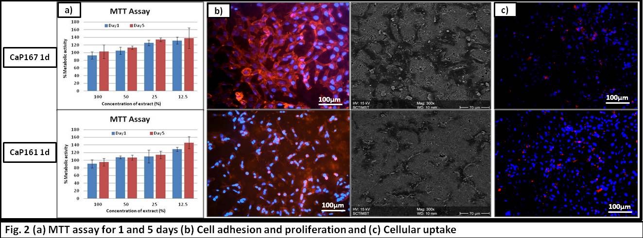
MTT assay confirms the non cytotoxic nature of the particles (Fig. 2a). Cell adhesion and proliferation on the surface of pelletized particles confirms the biocompatibility (Fig. 2b). Cellular uptake studies confirm the potential of the particles to act as therapeutic delivery agents.
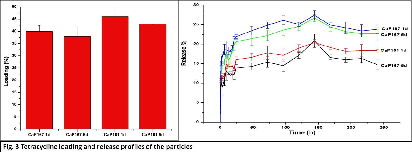
The loading and release profiles reveal that particles with a CaP ratio of 1.61 show a higher loading but a lesser release when compared to those with a CaP ratio of 1.67. Also smaller particles show a better loading and release profiles than their larger size counterparts.
Conclusion: In conclusion, a modified electrospraying process has been used to produce HA and CDHA particles in different size regimes. The particles do not elicit any toxicity and are readily taken up by the cells. Thus,electrosprayed CaP particles with controllable sizes are valid candidates for targeted delivery of therapeutic agents.The process opens up avenues for producing other ceramic particles as well.
Dr. H. K. Varma, Bioceramics Laboratory, SCTIMST
References:
[1] Sampath Kumar T. S., Yogeshwar Chakrapani V., Kumary T. V. “Process for producing calcium phosphate particles by electrospraying combined with sol-gel method” Patent Application no. 3071/CHE/2015 19.06.2015.
[2] Kettler K., Veltman K., Meent van de D., Wezel van A. & Hendriks A. J. “Cellular uptake of nanoparticles as determined by particle properties, experimental conditions , and cell type” Environmental Toxicology and Chemistry, 33(3), 481–492, 2014.