Introduction: Hydrogels made of natural polymers are interesting biomaterials because they simulate the aqueous environment of extracellular matrices and present high affinity with cells.[1] In particular, chitosan, a natural polysaccharide, exhibits excellent intrinsic properties such as biodegradability, biocompatibility and physiochemical characteristics offering unique opportunities for tissue engineering.[2] However, these soft hydrogels still present important challenges with respect to their clean fabrication (i.e., without using toxic chemicals) into precise structures featuring the appropriate mechanical resistance. In this work we propose the solvent-cast 3D printing technique to obtain precise and biocompatible three-dimensional (3D) chitosan scaffolds.
Materials and methods: Chitosan (degree of deacetylation = 90%, molecular weight = 207 kDa) used in this study was supplied by Biolog GmbH (Germany). 8 w/v % chitosan dissolved in mixed acids: acetic acid, lactic acid and citric acid (mole fraction: 71.1%, 27.3% and 1.6%, respectively) was prepared as the printable ink used for the fabrication. The solvent-cast 3D printing was developed to fabricate chitosan scaffolds at room temperature. The setup consists of a computer-controlled translation stage and a three-axis positioning platform. Chitosan scaffolds were produced by extruding the ink filaments on the plate in a layer–by-layer manner, which was followed by the filament solidification through solvent evaporation. Physical hydrogel scaffolds were obtained by neutralization in a sodium hydroxide solution. The tensile properties of chitosan scaffolds were examined using a MTS-Insight machine. Indirect toxicity (on extracts) of the scaffold was tested with L929 fibroblasts using the alamar blue test. The ability of cells to adhere and grow on material was also evaluated by alamar blue and live/dead assay after 1, 4 and 7 days.
Results and discussion: Chitosan scaffolds with a well-controlled pore size (minimum pore size as small as ~120 µm) and interconnected multilayer microfiber networks were achieved. Printed chitosan scaffolds (Fig.1) were self-supporting and can easily be manipulated and deformed with a tweezer without breaking. Chitosan scaffolds demonstrated relatively high values of elongation at break (around 110%) and Young’s modulus (around 22 MPa), fulfilling the demands for certain targeted tissues like cardiac muscles (i.e., 500 kPa). Our cell culture study showed that extracts from chitosan scaffolds were not toxic. Moreover cells adhered and grew on the scaffolds and the chitosan microfibers were almost fully covered by cells after 7 days (Fig.2).
Conclusion: We have successfully fabricated strong 3D chitosan scaffolds by solvent-cast 3D printing and observed a significant cell growth after 7 days. 3D printing truly offers a low-cost, scalable, highly flexible and efficient manufacturing technique for tissue engineering. Our strategy may also be applicable to other soft hydrogels based on self-assembling biomolecules such as collagen. Hybrid complex topologies with several soft hydrogels and tunable mechanical properties of scaffolds will be further developed to significantly advance 3D tissue-like structures.
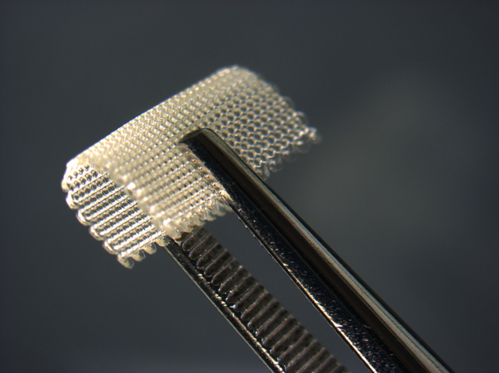
Figure 1. Optical microscopy image of 10-layer chitosan scaffolds folded using a tweezer

Figure 2. Optical fluorescence microscopy image of fibroblasts grew on scaffolds during 7 days
References:
[1] Fedorovich, N. E.; Alblas, J.; de Wijn, J. R.; Hennink, W. E.; Verbout, A. J.; Dhert, W. J., Hydrogels as extracellular matrices for skeletal tissue engineering: state-of-the-art and novel application in organ printing. Tissue engineering 2007, 13, (8), 1905-1925.
[2] Croisier, F.; Jérôme, C., Chitosan-based biomaterials for tissue engineering. European Polymer Journal 2013, 49, (4), 780-792.