There is a close correlation between vascularization and bone formation in endochondral ossification as maximum extent of bone formation follows maximum levels of VEGF expression. This suggests that osteogenesis and vascularization may be coupled by spatiotemporal regulation of paracrine signaling in which the invading vascular endothelial cells secrete osteogenic morphogens to stimulate cell differentiation and bone formation. The objective of this work was to investigate the effect of timed and localized release of BMP-2 and VEGF on the extent of osteogenic and vasculogenic differentiation of human mesenchymal stem cells (hMSCs) and endothelial colony-forming cells (ECFCs) in a patterned hydrogel co-culture system.
Methods: Polyethylene glycol (PEG) polymer chain-extended with lactide (L) and glycolide (G) segments or GL-PEG-GL macromers with different L/G feed molar ratios and PEG molecular weights were synthesized by sequential ring opening polymerization. The macromers were functionalized by reaction with disuccinimidyl carbonate (DCS). The macromers were self-assembled to nanogels (NGs) by dialysis. BMP-2 and VEGF proteins were grafted to the NGs by succinimide-amine reaction BMP2-NG and VEGF-NG). Size distribution of the NGs was measured by dynamic light scattering. NGs degradation and protein release was measured by incubation in PBS at 37°C. The protein release from the NGs was measured by ELISA. A 3D co-culture system with localized delivery of BMP-2 and VEGF was developed with a matrix of lactide-chain-extended PEG acrylate (SPELA) hydrogel and microchannels of gelatin methacrylate (GelMA) as shown in Figure 1. hMSCs and BMP2-NG were encapsulated in the RGD-functionalized SPELA and a mixture of hMSCs+hECFCs with VEGF-NG were encapsulated in the channels. The 3D co-culture system was cultivated in osteogenic-vasculogenic medium for 21 days. At each time point, the co-cultures were evaluated for osteogenesis and vasculogenesis by biochemical, mRNA, and protein analysis.
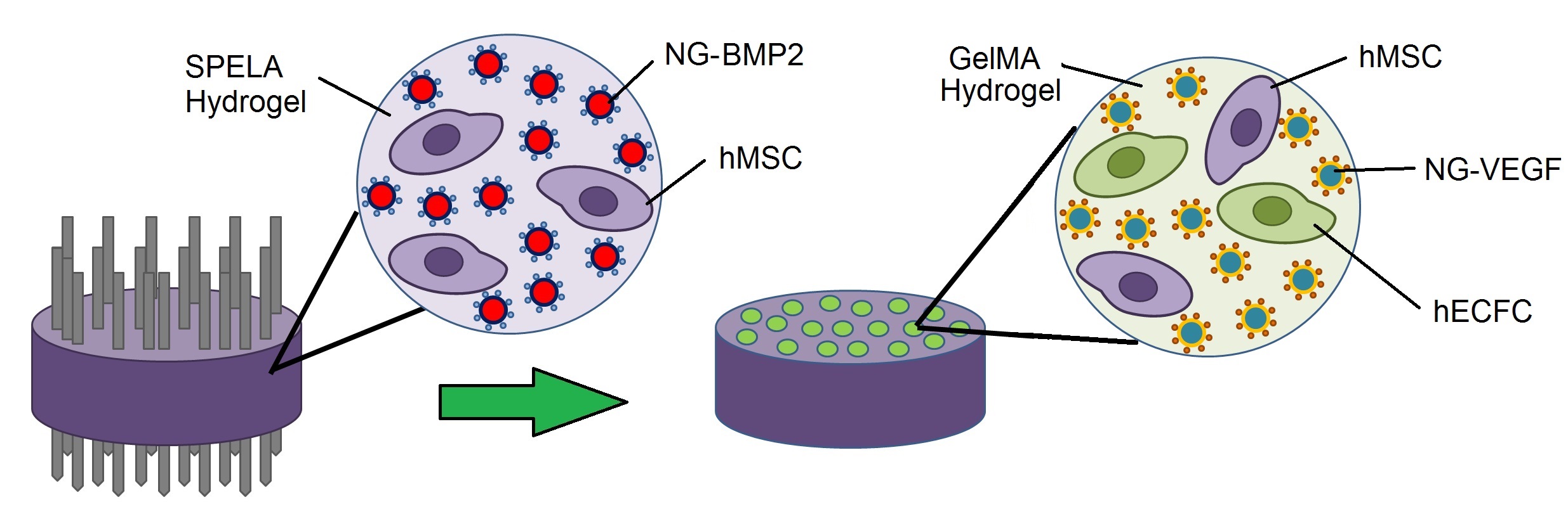
Figure 1. Schematic diagram of vasculogenic GelMA microchannels in osteogenic SPELA gel for patterned constructs.
ALP activity of hMSCs in un-patterned osteogenic constructs with BMP2 or BMP2-NGs (dashed curves) increased significantly from day 7 to 14, reached a maximum after 14 days and decreased from day 14 to 21. The patterned constructs (solid lines) had higher ALP activity than their corresponding un-patterned osteogenic constructs (dashed lines). The patterned constructs with NG-BMP2/NG-VEGF (blue curve) and without BMP2/VEGF (green curve) had the highest and lowest ALP activity with 6100±500 and 2000±300 IU/mg DNA, respectively. CD31 protein expression for the patterned construct without BMP2/VEGF (green curve) did not increase significantly with time (Figure 2). CD31 expression of the un-patterned vasculogenic constructs with VEGF (dashed pink) or NG-VEGF (dashed light blue) increased with time but the CD31 expression for the NG-VEGF construct was significantly higher than that of VEGF. The patterned constructs (solid lines) had higher CD31 expression than the un-patterned vasculogenic constructs (dashed lines) for all incubation times. For all time points, CD31 expression of patterned construct with NG-BMP2/NG-VEGF was higher than the other groups.
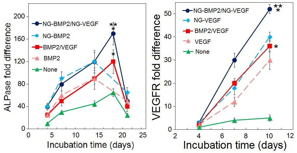
Figure 2. ALPase and VEGF receptor mRNA expression of hMSCs and hECFCs in the patterned constructs.
Conclusions: The extent of osteogenic and vasculogenic differentiation of hMSCs and hECFCs was higher in patterned compared to un-patterned constructs. Further, timed-release of VEGF and BMP2 from the NGs in the patterned constructs significantly enhanced osteogenic differentiation of MSCs and vasculogenic differentiation of hMSCs+hECFCs compared with direct addition of VEGF and BMP2.
National Science Foundation (USA) grants IIP150024 and CBET1403545; National Institutes of Health (USA) grant AR063745