Introduction: When a biomaterial is inserted into the cardiovascular system, within seconds of blood-material contact, the surface is covered by a layer of proteins, where the composition, relative concentration, conformation, and orientation play a key role in determining the fate of the material[1]. In this study, protein adsorption onto polyurethane (PU) films with different surface functional groups and surface characteristics (wettability, surface energy, chemistry, etc.) were examined in order to understand the interaction between the proteins and the surface.
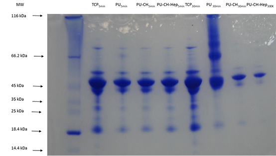
Materials and Methods: PU was synthesized by condensation polymerization of polypropylene ethylene glycol (PPEG) and toluene diisocyanate (TDI) without adding any other ingredient. Surfaces of PU films were modified by a step wise process via oxygen plasma activation, acrylamide treatment and then chitosan (CH) grafting (PU-CH). Also, immobilization of heparin (Hep) on chitosan grafted PU films were carried out as multilayer surface modification (PU-CH-Hep). Modified surface films were characterized by ESCA, AFM and goniometer[2]. Platelet poor human plasma was incubated on samples at different time intervals of 5 and 30 min, after the removal of adhered plasma layer, proteins were characterized by SDS-page, Bradford assay and SEM. The endocrinic cytotoxicity of PU film samples was examined by MTT assay by using HEK293 cells.
Result and Discussion: The surface atomic composition of PU film has changed after CH and Hep immobilization. Chitosan grafting caused an increase in nitrogen content (3.1% for PU and 4.8% for PU-CH) because of amine groups of chitosan. Sulfur, an investigative atom for heparin, was found on PU-CH-Hep (0.4%) which indicates presence of Hep on PU-CH-Hep samples. PU-CH samples, which had the highest roughness with brush-like morphology, exhibited lowest protein adsorption. Modifications improved surface wettability. PU-CH-Hep samples demonstrated highest hydrophilicity with lowest contact angle (34o) and exhibited protein adsorption in the early stage of the surface-plasma interaction, with minimal aggregation and no thrombus like formation. No toxic effect was observed for PU showing complete polymerization with no remaining monomer due to high degree of polymerization.
Table 1. Surface atomic compositions and water contact angles (%) of PU, PU-CH and PU-CH-Hep films


Figure 1. Electrophoresis profiles of binding strength of low MW plasma proteins
Conclusion: Polyurethanes modified with chitosan and heparin grafting demonstrated different adsorptions for plasma proteins due to variations on the chemistry, functionality, topography and wettability of the surfaces.
We gratefully acknowledge the support to BIOMATEN through the Ministry of Development funds and by METU; Characterization analyses were carried out by Central Lab of METU
References:
[1] Aksoy E.A., Hasirci V., Hasirci N., Motta A., Fedel M., Migliaresi C., Journal of Bioactive and Compatible Polymers, 23: pp.505-519, 2008.
[2] Kara F., Aksoy E.A., Yuksekdag Z., Hasirci N., Aksoy S., Carbohydrate Polymers, 112: pp.39–47, 2014