Introduction: Patterned hydrogels and polymer scaffolds have attracted attention as platforms for directed cell growth due to the significant impacts cell alignment has on tissue regeneration, mechanical properties and various other cell behaviours[1],[2]. However, most currently reported platforms require lengthy micropatterning steps[3]-[10], limiting their clinical applicability in vitro. Here, we propose an injectable hydrogel platform with magnetically aligned cellulose nanocrystals (CNCs) that offers potential applicability as a platform for cell growth both in vitro and in vivo.
Materials and Methods: Injectable poly (oligo ethylene glycol) methacrylate (POEGMA) hydrogels were synthesized and prepared as described previously[11]. High aspect ratio CNCs were made via sulfuric acid catalyzed hydrolysis of cotton and were physically incorporated into POEGMA hydrogels, similar to a previous report[12]. A 0.56T rare earth magnet was used for in situ alignment of CNCs within nanocomposite hydrogels directly following injection into molds. Small angle X-ray scattering (SAXS) measurements were performed in to quantify CNC alignment within hydrogel samples. Bulk hydrogel properties were characterized via swelling and rheology measurements, while cryo-TEM was used to observe the CNC distribution within hydrogels. Cell adhesion experiments were performed on C2C12 myoblast cells, and observed using fluorescence microscopy (2D) or confocal fluorescence microscopy (3D).
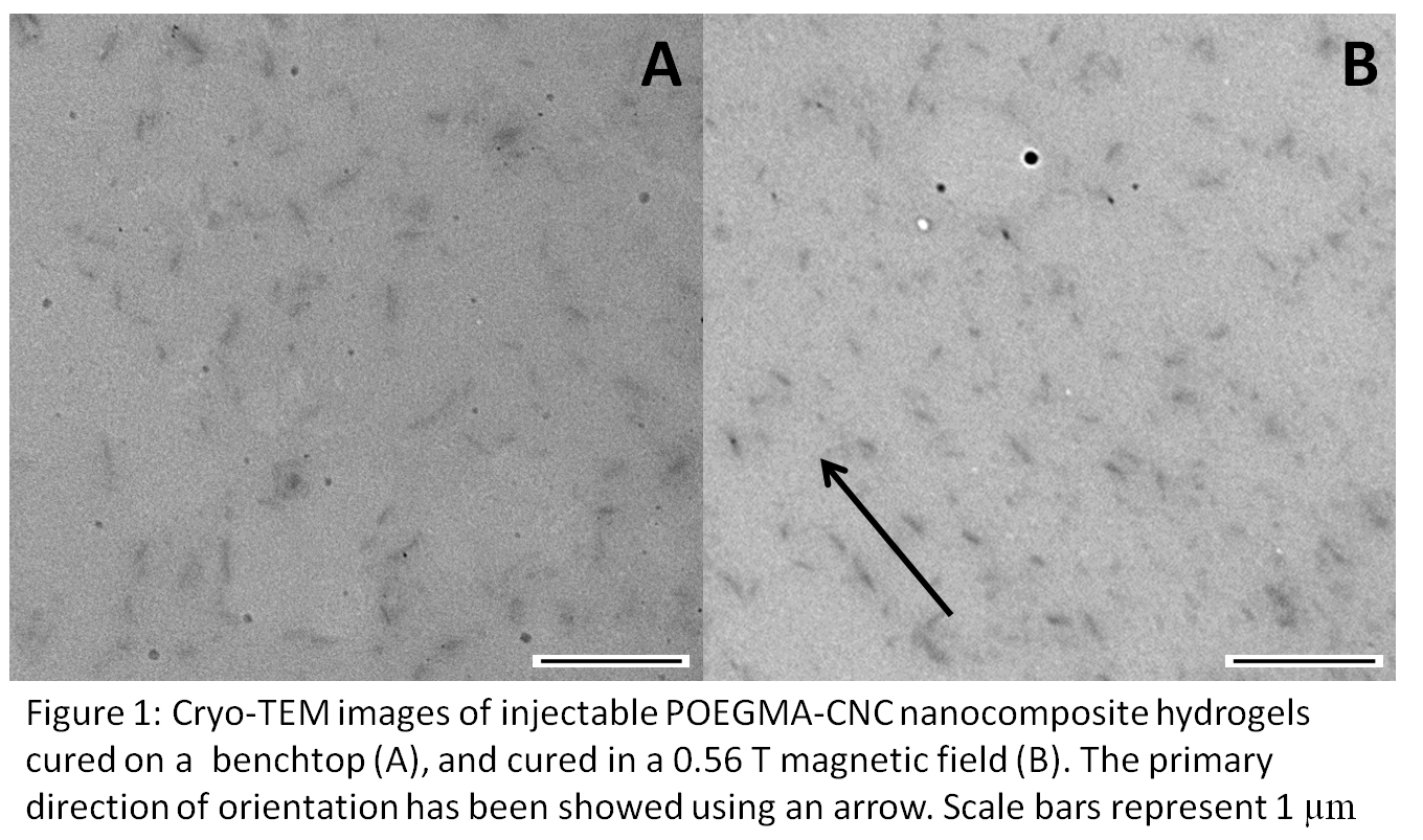
Results: Significant CNC alignment within nanocomposite POEGMA hydrogels was achieved using a 0.56T rare earth magnet, as evidenced by SAXS data and cryo-TEM images of internal hydrogel sections (Figure 1).
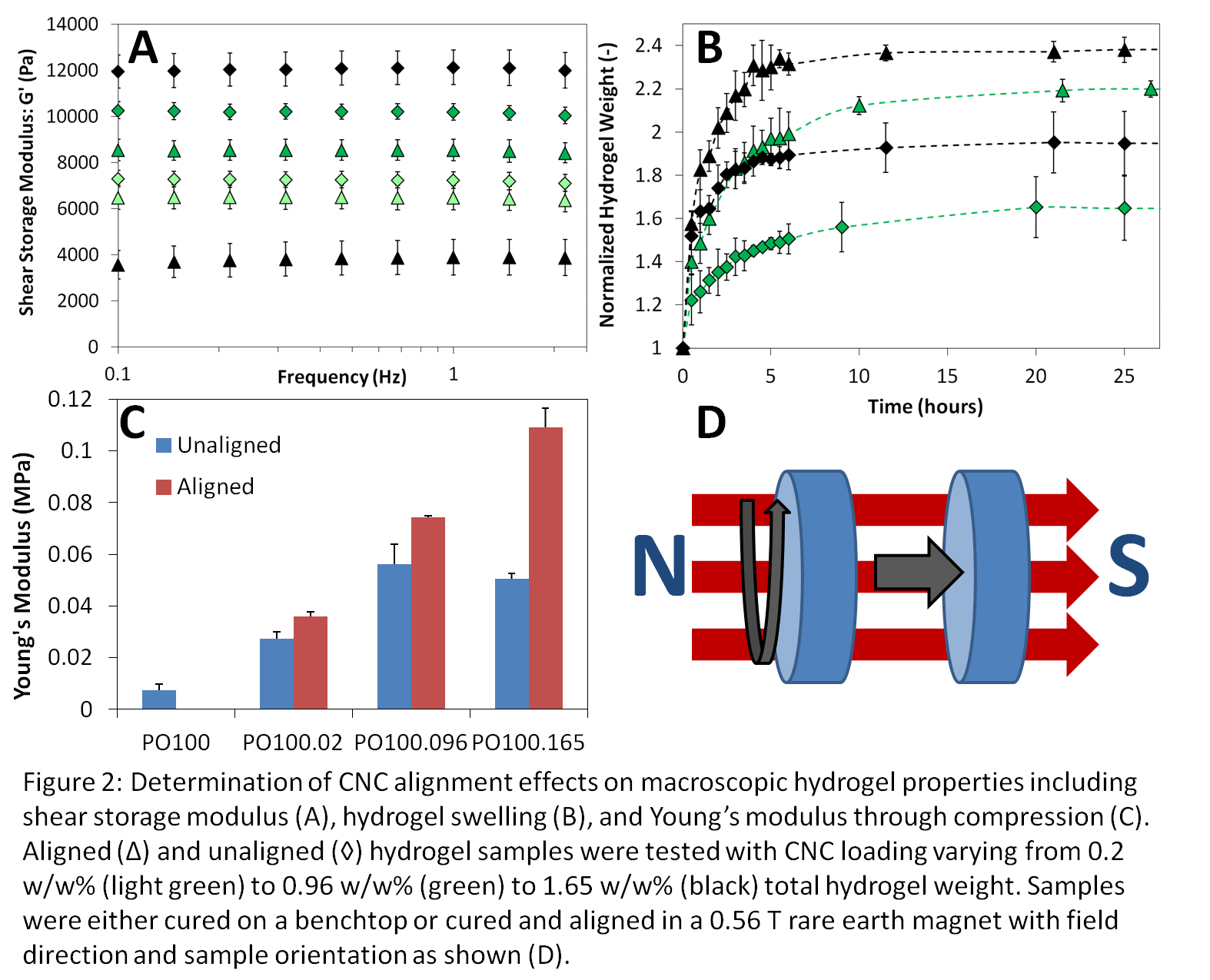
Alignment of CNCs within POEGMA hydrogels leads to a decrease in hydrogel swelling, along with a decrease in shear storage modulus but an increase in compressive modulus (Figure 2).
Increasing the CNC loading within the hydrogels increased the level of cell adhesion, beneficial for tissue engineering scaffolds. Further, C2C12 myoblast adhesion was observed for unaligned and aligned hydrogel samples having similar CNC loading, with preliminary evidence of enhanced alignment in the magnetically aligned hydrogels.
Discussion: The diamagnetic susceptibility of CNCs has been well documented, persuading CNCs to align effectively perpendicular to a magnetic field. Previous studies have focused on aligning pure suspensions of CNCs in extremely high magnetic fields over many hours[13],[14]. To our knowledge, this is the first experimental evidence of significant CNC alignment achieved in a relatively low magnetic field in a matter of minutes. Moreover, this is the first experimental evidence of CNCs aligning during in situ gelation of a chemically crosslinked polymer hydrogel. TEM and SAXS observations support the successful magnetic alignment of CNCs, giving rise to hydrogels with tailorable swelling and rheological properties with potential use in numerous tissue engineering applications.
Conclusion: We have demonstrated successful in situ magnetic alignment of rod-like CNCs within a nanocomposite POEGMA hydrogel matrix, as confirmed through both TEM and SAXS observations. These hydrogels exhibit tailorable swelling and rheological properties due to both CNC loading and the presence of an external magnetic field. Hydrogels with aligned CNCs showed an increase in the alignment of adhered C2C12 myoblasts, suggesting exceptional opportunities for their use in tissue engineering applications.
National Sciences and Engineering Research Council of Canada; NSERC CREATE-IDEM
References:
[1] Li, Y. et al. Engineering cell alignment in vitro. Biotechnol. Adv. 32, 347–365 (2014)
[2] Dugan, J. M., Collins, R. F., Gough, J. E. & Eichhorn, S. J. Oriented surfaces of adsorbed cellulose nanowhiskers promote skeletal muscle myogenesis. Acta Biomater. 9, 4707–15 (2013)
[3] Chan, V. et al. Directed cell growth and alignment on protein-patterned 3D hydrogels with stereolithography. Virtual Phys. Prototyp. 7, 219–228 (2012)
[4] Zhu, B. et al. Nanotopographical guidance of C6 glioma cell alignment and oriented growth. Biomaterials 25, 4215–4223 (2004)
[5] Breukers, R. D. et al. Creating conductive structures for cell growth: Growth and alignment of myogenic cell types on polythiophenes. J. Biomed. Mater. Res. - Part A 95, 256–268 (2010)
[6] Aubin, H. et al. Directed 3D cell alignment and elongation in microengineered hydrogels. Biomaterials 31, 6941–6951 (2010)
[7] Krsko, P. et al. Length-scale mediated adhesion and directed growth of neural cells by surface-patterned poly(ethylene glycol) hydrogels. Biomaterials 30, 721–729 (2009)
[8] Lee, S. H., Moon, J. J. & West, J. L. Three-dimensional micropatterning of bioactive hydrogels via two-photon laser scanning photolithography for guided 3D cell migration. Biomaterials 29, 2962–2968 (2008)
[9] DeLong, S. a., Gobin, A. S. & West, J. L. Covalent immobilization of RGDS on hydrogel surfaces to direct cell alignment and migration. J. Control. Release 109, 139–148 (2005)
[10] Guarnieri, D. et al. Covalently immobilized RGD gradient on PEG hydrogel scaffold influences cell migration parameters. Acta Biomater. 6, 2532–2539 (2010)
[11] Smeets, N. M. B., Bakaic, E., Patenaude, M. & Hoare, T. Injectable poly(oligoethylene glycol methacrylate)-based hydrogels with tunable phase transition behaviours: physicochemical and biological responses. Acta Biomater. 10, 4143–55 (2014)
[12] Yang, X., Bakaic, E., Hoare, T. & Cranston, E. D. Injectable polysaccharide hydrogels reinforced with cellulose nanocrystals: morphology, rheology, degradation, and cytotoxicity. Biomacromolecules 14, 4447–55 (2013)
[13] Pullawan, T., Wilkinson, A. N. & Eichhorn, S. J. Influence of magnetic field alignment of cellulose whiskers on the mechanics of all-cellulose nanocomposites. Biomacromolecules 13, 2528–36 (2012)
[14] Abitbol, T. & Cranston, E. D. in Handbook of Green Materials 79–103 (2012)